How to start getting out of depression, ADHD, OCD, anxiety and...

George Ferman@Helios_Movement
163 views
Jul 21, 2026
~17 min read
Advertisement
1
How to start getting out of depression, ADHD, OCD, anxiety and panic disorders by utilizing GABA.
Thread🧵
Thread🧵
2
It's George.
The brain contains a vast neural architecture where billions of cells continuously exchange information.
This complex signaling is driven by neurotransmitters, the molecular messengers responsible for cellular communication.
So by attempting to decode neurotransmitters, we are attempting to map the specific language the brain uses to send instructions and process our thoughts and emotions.
Overall, in case you are new here, neurotransmitters are chemical substances that neurons release to send signals across synapses, the tiny gaps between nerve cells.
These molecules are categorized based on their effects.
We have:
-Excitatory neurotransmitters that increase the likelihood of a neuron firing an action potential.
-Inhibitory neurotransmitters that decrease the likelihood of a neuron firing, providing a calming effect.
-Modulatory neurotransmitters that influence the overall activity of groups of neurons, modulating their function over time.
The brain contains a vast neural architecture where billions of cells continuously exchange information.
This complex signaling is driven by neurotransmitters, the molecular messengers responsible for cellular communication.
So by attempting to decode neurotransmitters, we are attempting to map the specific language the brain uses to send instructions and process our thoughts and emotions.
Overall, in case you are new here, neurotransmitters are chemical substances that neurons release to send signals across synapses, the tiny gaps between nerve cells.
These molecules are categorized based on their effects.
We have:
-Excitatory neurotransmitters that increase the likelihood of a neuron firing an action potential.
-Inhibitory neurotransmitters that decrease the likelihood of a neuron firing, providing a calming effect.
-Modulatory neurotransmitters that influence the overall activity of groups of neurons, modulating their function over time.
3
GABA is the one that counterbalances the excitatory actions of glutamate and helps maintain the excitation-inhibition (E/I) balance, which is essential for proper brain function.
Reduced GABAergic tone frequently disrupts this equilibrium and leads to neuronal hyperexcitability (and a widespread over-excitation in different brain regions overall), which is a state implicated in numerous neuropsychiatric and neurological disorders.
This is why if you experience symptoms and conditions such as:
-Generalized anxiety disorder
-Panic disorder
-Racing thoughts
-Sleep-onset insomnia and fragmented sleep architecture
-Muscle tension and physical restlessness
-Heightened startle response
-Bipolar disorder
-ADHD
-Emotional reactivity / irritability
-Difficulty focusing / sensory overload
-Major Depressive Disorder
-Autism Spectrum Disorder (ASD)
-Schizophrenia
-Epilepsy (various syndromes)
-Obsessive-Compulsive Disorder (OCD)
-Chronic pain and central sensitization
-Fibromyalgia
-Alcohol withdrawal syndrome
-Post-traumatic stress disorder (PTSD)
then you likely need to explore GABA.
Reduced GABAergic tone frequently disrupts this equilibrium and leads to neuronal hyperexcitability (and a widespread over-excitation in different brain regions overall), which is a state implicated in numerous neuropsychiatric and neurological disorders.
This is why if you experience symptoms and conditions such as:
-Generalized anxiety disorder
-Panic disorder
-Racing thoughts
-Sleep-onset insomnia and fragmented sleep architecture
-Muscle tension and physical restlessness
-Heightened startle response
-Bipolar disorder
-ADHD
-Emotional reactivity / irritability
-Difficulty focusing / sensory overload
-Major Depressive Disorder
-Autism Spectrum Disorder (ASD)
-Schizophrenia
-Epilepsy (various syndromes)
-Obsessive-Compulsive Disorder (OCD)
-Chronic pain and central sensitization
-Fibromyalgia
-Alcohol withdrawal syndrome
-Post-traumatic stress disorder (PTSD)
then you likely need to explore GABA.
4
Of course, low GABA tone/dysfunctional GABA signaling is rarely the sole cause of any of these and it usually interacts with inflammation, genetics, stress, and a few other factors besides glutamate excess.
But many of these symptoms/conditions improve when GABA tone is supported through lifestyle, nutrition, or targeted therapies.
So it’s a valuable puzzle piece towards resolving any of these.
Overall, if GABA was irrelevant, benzodiazepines wouldn’t be so popular and abused or GABAB receptor-mediated inhibition is wouldn’t be so dysregulated in depressed adolescents (especially the ones with histories of suicidal behavior).
But many of these symptoms/conditions improve when GABA tone is supported through lifestyle, nutrition, or targeted therapies.
So it’s a valuable puzzle piece towards resolving any of these.
Overall, if GABA was irrelevant, benzodiazepines wouldn’t be so popular and abused or GABAB receptor-mediated inhibition is wouldn’t be so dysregulated in depressed adolescents (especially the ones with histories of suicidal behavior).
5
Now GABA (γ-aminobutyric acid) is a non-proteinogenic amino acid(*) that serves as the primary inhibitory neurotransmitter in the mammalian CNS and was isolated and identified as a major component of brain tissue by Eugene Roberts and Sam Frankel around 1950.
(*) Proteinogenic amino acids (the 20 standard ones + a few others like selenocysteine) are used by ribosomes to build proteins according to the genetic code.
GABA is non-proteinogenic, so it is not incorporated into proteins during protein synthesis.
It has a different structure (the amino group is on the gamma carbon instead of the alpha carbon), so it cannot be used in the standard translation process.
So GABA functions almost exclusively as a signaling molecule (neurotransmitter) and metabolic intermediate, not as a structural building block of proteins.
This is why it is rarely just called an “amino acid” without the qualifier “non-proteinogenic” in scientific literature.
It mediates inhibitory neurotransmission at roughly 20–50% of all synapses in the brain with particularly dense representation in (*)the cerebral cortex, hippocampus, amygdala, basal ganglia, and spinal cord.
You may find the range 30%-40% in some textbooks, but usually, most agree that’s roughly 20%-50%.
(*) Meta-analyses of magnetic resonance spectroscopy (MRS) studies show significantly reduced GABA concentrations in these exact key brain regions in individuals with active major depression, anxiety disorders, panic disorders and ASD.
(*) Proteinogenic amino acids (the 20 standard ones + a few others like selenocysteine) are used by ribosomes to build proteins according to the genetic code.
GABA is non-proteinogenic, so it is not incorporated into proteins during protein synthesis.
It has a different structure (the amino group is on the gamma carbon instead of the alpha carbon), so it cannot be used in the standard translation process.
So GABA functions almost exclusively as a signaling molecule (neurotransmitter) and metabolic intermediate, not as a structural building block of proteins.
This is why it is rarely just called an “amino acid” without the qualifier “non-proteinogenic” in scientific literature.
It mediates inhibitory neurotransmission at roughly 20–50% of all synapses in the brain with particularly dense representation in (*)the cerebral cortex, hippocampus, amygdala, basal ganglia, and spinal cord.
You may find the range 30%-40% in some textbooks, but usually, most agree that’s roughly 20%-50%.
(*) Meta-analyses of magnetic resonance spectroscopy (MRS) studies show significantly reduced GABA concentrations in these exact key brain regions in individuals with active major depression, anxiety disorders, panic disorders and ASD.
6
GABAergic neurons and terminals are also dense in several other regions worth naming individually, since each maps onto a specific function:
-The thalamus (relays sensory and motor information to the cortex).
-The hypothalamus (regulates hormones, body temperature, hunger, and the stress response).
-The brainstem (controls breathing, heart rate, sleep, and other vital automatic functions).
-The cerebellum (coordination, balance, posture, and motor learning)
-The olfactory bulb (processes smell).
-The retina (processes visual information before it reaches the brain).
-The superior colliculus (coordinates eye and head movements in response to visual stimuli).
-The inferior colliculus (processes auditory information).
-The vestibular nuclei (maintain balance and spatial orientation).
-The suprachiasmatic nucleus or SCN (controls circadian rhythm and the sleep-wake cycle).
This widespread distribution is why GABA touches nearly every major aspect of brain function, not just mood and anxiety:
-Attention
-Memory
-Movement
-Sensory processing
-Pain perception
-Motor coordination
-The sleep-wake cycle
….are all downstream of it.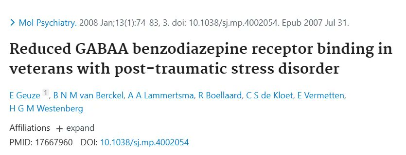
-The thalamus (relays sensory and motor information to the cortex).
-The hypothalamus (regulates hormones, body temperature, hunger, and the stress response).
-The brainstem (controls breathing, heart rate, sleep, and other vital automatic functions).
-The cerebellum (coordination, balance, posture, and motor learning)
-The olfactory bulb (processes smell).
-The retina (processes visual information before it reaches the brain).
-The superior colliculus (coordinates eye and head movements in response to visual stimuli).
-The inferior colliculus (processes auditory information).
-The vestibular nuclei (maintain balance and spatial orientation).
-The suprachiasmatic nucleus or SCN (controls circadian rhythm and the sleep-wake cycle).
This widespread distribution is why GABA touches nearly every major aspect of brain function, not just mood and anxiety:
-Attention
-Memory
-Movement
-Sensory processing
-Pain perception
-Motor coordination
-The sleep-wake cycle
….are all downstream of it.
7
In MDD for example postmortem studies show decreased expression of GAD67 and parvalbumin in prefrontal interneurons (we will talk about both of these in a moment).
Low GABA basically impairs prefrontal regulation of limbic structures, so the person has a very hard time getting out of negative thought loops.
Notably, successful antidepressant treatment (pharmacological or neuromodulatory) often normalizes cortical GABA levels, suggesting state-dependent rather than purely trait-related changes.
In schizophrenia, postmortem and imaging data also reveal deficits in GABAergic interneurons (particularly parvalbumin-positive cells that we’ll briefly talk about in a moment) and altered GAD expression, contributing to impaired gamma oscillations and sensory gating and in epilepsy, loss of inhibitory tone, whether through reduced GAD activity, interneuron loss, or chloride homeostasis disruption, lowers the seizure threshold.
Also, in generalized anxiety disorder, positron emission tomography (PET) studies demonstrate reduced benzodiazepine-binding site density in the amygdala, prefrontal cortex, and insula, consistent with diminished GABAA receptor availability and benzodiazepine sensitivity.
There also seems to be reduced GABAA-benzodiazepine binding sites in the insular cortex of individuals with panic disorder and PTSD.
All of these make sense even if we didn’t have any studies ont them, since dense GABAergic innervation of the amygdala, prefrontal cortex, and hippocampus enables top-down control of fear and stress responses.
So the hypo-inhibition experienced in MDD, GAD and so on, permits excessive amygdala reactivity to neutral or mildly threatening stimuli for example, producing persistent worry, autonomic hyperarousal, and even panic attacks.
Low GABA basically impairs prefrontal regulation of limbic structures, so the person has a very hard time getting out of negative thought loops.
Notably, successful antidepressant treatment (pharmacological or neuromodulatory) often normalizes cortical GABA levels, suggesting state-dependent rather than purely trait-related changes.
In schizophrenia, postmortem and imaging data also reveal deficits in GABAergic interneurons (particularly parvalbumin-positive cells that we’ll briefly talk about in a moment) and altered GAD expression, contributing to impaired gamma oscillations and sensory gating and in epilepsy, loss of inhibitory tone, whether through reduced GAD activity, interneuron loss, or chloride homeostasis disruption, lowers the seizure threshold.
Also, in generalized anxiety disorder, positron emission tomography (PET) studies demonstrate reduced benzodiazepine-binding site density in the amygdala, prefrontal cortex, and insula, consistent with diminished GABAA receptor availability and benzodiazepine sensitivity.
There also seems to be reduced GABAA-benzodiazepine binding sites in the insular cortex of individuals with panic disorder and PTSD.
All of these make sense even if we didn’t have any studies ont them, since dense GABAergic innervation of the amygdala, prefrontal cortex, and hippocampus enables top-down control of fear and stress responses.
So the hypo-inhibition experienced in MDD, GAD and so on, permits excessive amygdala reactivity to neutral or mildly threatening stimuli for example, producing persistent worry, autonomic hyperarousal, and even panic attacks.
8
At the cellular level, GABA released into the synaptic cleft binds primarily to two receptor classes:
Ionotropic GABAA receptors
These are ligand-gated ion channels (pentameric structures made of five protein subunits).
When GABA binds, they open and allow chloride ions (Cl⁻) to flow into the neuron.
In mature neurons, this causes hyperpolarization (the inside of the cell becomes more negative), making it harder for the neuron to fire.
To put a number on that: a neuron typically rests at around −70 mV.
Once chloride rushes in, the membrane potential drops further, often to around −80 mV or lower, while the threshold needed to fire an action potential sits at roughly −55 mV.
So a hyperpolarized neuron needs a much bigger excitatory push just to get back to where it can fire at all.
This is what “raising the bar” for glutamate-driven signals actually looks like in voltage terms.
They can also produce shunting inhibition, which dampens excitatory signals even without major voltage change, and by keeping the membrane well below threshold for long enough, GABA effectively prevents the voltage sensors on nearby sodium channels from activating in the first place, blocking the propagation of a signal before it can spread.
Some of their key features include:
Phasic inhibition: Fast, brief bursts at synapses (mainly synaptic receptors).
Tonic inhibition: Steady, low-level background inhibition (extrasynaptic receptors, often containing δ subunits).
There are 19 different subunits (α1–6, β1–4, γ1–3, δ, etc. with rarer ones like ε, θ, π, and ρ1–3 also part of the full family).
The most common combination is α1β2γ2. Subunit composition determines:
α1 → Strong sedation and anticonvulsant effects (target of many sleeping pills).
α2 & α3 → Anxiolytic (anti-anxiety) and muscle-relaxant effects.
α5 → Contributes more to cognitive processes, particularly learning and memory, rather than sedation or anxiolysis.
δ-containing → Tonic inhibition, seizure threshold, and sensitivity to alcohol/neurosteroids. These δ subunits typically pair specifically with α4 or α6 subunits.
Benzodiazepines, barbiturates, alcohol, and neurosteroids (like allopregnanolone) act here as positive allosteric modulators, enhancing GABA’s effect.
Ionotropic GABAA receptors
These are ligand-gated ion channels (pentameric structures made of five protein subunits).
When GABA binds, they open and allow chloride ions (Cl⁻) to flow into the neuron.
In mature neurons, this causes hyperpolarization (the inside of the cell becomes more negative), making it harder for the neuron to fire.
To put a number on that: a neuron typically rests at around −70 mV.
Once chloride rushes in, the membrane potential drops further, often to around −80 mV or lower, while the threshold needed to fire an action potential sits at roughly −55 mV.
So a hyperpolarized neuron needs a much bigger excitatory push just to get back to where it can fire at all.
This is what “raising the bar” for glutamate-driven signals actually looks like in voltage terms.
They can also produce shunting inhibition, which dampens excitatory signals even without major voltage change, and by keeping the membrane well below threshold for long enough, GABA effectively prevents the voltage sensors on nearby sodium channels from activating in the first place, blocking the propagation of a signal before it can spread.
Some of their key features include:
Phasic inhibition: Fast, brief bursts at synapses (mainly synaptic receptors).
Tonic inhibition: Steady, low-level background inhibition (extrasynaptic receptors, often containing δ subunits).
There are 19 different subunits (α1–6, β1–4, γ1–3, δ, etc. with rarer ones like ε, θ, π, and ρ1–3 also part of the full family).
The most common combination is α1β2γ2. Subunit composition determines:
α1 → Strong sedation and anticonvulsant effects (target of many sleeping pills).
α2 & α3 → Anxiolytic (anti-anxiety) and muscle-relaxant effects.
α5 → Contributes more to cognitive processes, particularly learning and memory, rather than sedation or anxiolysis.
δ-containing → Tonic inhibition, seizure threshold, and sensitivity to alcohol/neurosteroids. These δ subunits typically pair specifically with α4 or α6 subunits.
Benzodiazepines, barbiturates, alcohol, and neurosteroids (like allopregnanolone) act here as positive allosteric modulators, enhancing GABA’s effect.
9
Metabotropic GABAB receptors.
These are G-protein-coupled receptors (metabotropic), working more slowly but with longer-lasting effects.
They exist as heterodimers (GABAB1 + GABAB2).
When GABA binds to GABAB receptors, it triggers two main effects depending on where the receptor is located:
1. Presynaptic GABAB receptors (on the sending neuron)
These act as autoreceptors or heteroreceptors.
Activation → inhibits voltage-gated calcium channels (specifically N-type and P/Q-type Ca²⁺ channels).
Result: Less calcium enters the presynaptic terminal when an action potential arrives.
Because calcium is required for neurotransmitter vesicles to fuse and release their contents, less calcium = less neurotransmitter release.
This includes:
Reduced release of glutamate (excitatory) → overall calming effect.
Reduced release of GABA itself (negative feedback on the inhibitory neuron).
This is a form of presynaptic inhibition that dampens both excitatory and inhibitory signaling over a longer timescale.
2. Postsynaptic GABAB receptors (on the receiving neuron)
Activation couples to G-proteins that open inwardly rectifying potassium channels (GIRK channels).
Potassium ions (K⁺) flow out of the cell.
Result: The inside of the neuron becomes more negative → slow hyperpolarization.
This makes the postsynaptic neuron less excitable for a longer period (hundreds of milliseconds to seconds), producing a slow, sustained inhibitory effect.
So GABAB receptors are important for:
Regulating network oscillations
Pain modulation
Mood stability
Preventing excessive excitation over longer time scales
Baclofen is a GABAB agonist used for muscle spasticity and sometimes alcohol dependence.
This variety of GABAA receptor subtypes allows the brain to fine-tune inhibition for different needs:
Fast, precise control (phasic)
Steady background calm (tonic)
Region-specific effects (amygdala for fear, hippocampus for memory, cortex for thinking)
These are G-protein-coupled receptors (metabotropic), working more slowly but with longer-lasting effects.
They exist as heterodimers (GABAB1 + GABAB2).
When GABA binds to GABAB receptors, it triggers two main effects depending on where the receptor is located:
1. Presynaptic GABAB receptors (on the sending neuron)
These act as autoreceptors or heteroreceptors.
Activation → inhibits voltage-gated calcium channels (specifically N-type and P/Q-type Ca²⁺ channels).
Result: Less calcium enters the presynaptic terminal when an action potential arrives.
Because calcium is required for neurotransmitter vesicles to fuse and release their contents, less calcium = less neurotransmitter release.
This includes:
Reduced release of glutamate (excitatory) → overall calming effect.
Reduced release of GABA itself (negative feedback on the inhibitory neuron).
This is a form of presynaptic inhibition that dampens both excitatory and inhibitory signaling over a longer timescale.
2. Postsynaptic GABAB receptors (on the receiving neuron)
Activation couples to G-proteins that open inwardly rectifying potassium channels (GIRK channels).
Potassium ions (K⁺) flow out of the cell.
Result: The inside of the neuron becomes more negative → slow hyperpolarization.
This makes the postsynaptic neuron less excitable for a longer period (hundreds of milliseconds to seconds), producing a slow, sustained inhibitory effect.
So GABAB receptors are important for:
Regulating network oscillations
Pain modulation
Mood stability
Preventing excessive excitation over longer time scales
Baclofen is a GABAB agonist used for muscle spasticity and sometimes alcohol dependence.
This variety of GABAA receptor subtypes allows the brain to fine-tune inhibition for different needs:
Fast, precise control (phasic)
Steady background calm (tonic)
Region-specific effects (amygdala for fear, hippocampus for memory, cortex for thinking)
10
This diversity also explains why drugs like benzodiazepines can be both helpful (anxiety relief) and problematic (sedation, tolerance, dependence, profoundly cooked brain etc).
It’s also worth flipping the picture for a second: this whole article has been about what happens when GABA tone is too low, but too much inhibition causes its own problems:
Excessive sedation
Reduced alertness
Slower reaction times
Impaired coordination (ataxia)
Cognitive slowing
Memory difficulties
Reduced motivation
The goal was never to max out GABA activity as if more is automatically better; it’s to keep the E/I balance in a healthy range, since inhibition that’s too strong is just as disruptive as inhibition that’s too weak.
What’s also interesting is that in the fetal and newborn brain, high intracellular chloride makes GABAA activation excitatory rather than inhibitory.
This “GABA switch” is crucial for brain development and disruptions here are linked to autism, epilepsy, and other neurodevelopmental conditions overall.
It’s also worth flipping the picture for a second: this whole article has been about what happens when GABA tone is too low, but too much inhibition causes its own problems:
Excessive sedation
Reduced alertness
Slower reaction times
Impaired coordination (ataxia)
Cognitive slowing
Memory difficulties
Reduced motivation
The goal was never to max out GABA activity as if more is automatically better; it’s to keep the E/I balance in a healthy range, since inhibition that’s too strong is just as disruptive as inhibition that’s too weak.
What’s also interesting is that in the fetal and newborn brain, high intracellular chloride makes GABAA activation excitatory rather than inhibitory.
This “GABA switch” is crucial for brain development and disruptions here are linked to autism, epilepsy, and other neurodevelopmental conditions overall.
11
Now, GABA is synthesized in a single enzymatic step from glutamate by the enzyme glutamate decarboxylase (GAD).
Two major isoforms exist, each with distinct localization and functional roles:
GAD67 (encoded by GAD1, ~67 kDa): Primarily cytosolic and constitutively active. It maintains basal GABA levels for tonic inhibition and is especially critical during brain development.
GAD65 (encoded by GAD2, ~65 kDa): Membrane-associated and often found in an inactive apo-form near synaptic vesicles. It is rapidly activated in response to neuronal activity and supports phasic, on-demand GABA release.
The decarboxylation reaction (Glutamate + H⁺ → GABA + CO₂) requires pyridoxal 5′-phosphate (P5P), the bioactive form of vitamin B6, as an essential cofactor.
Two major isoforms exist, each with distinct localization and functional roles:
GAD67 (encoded by GAD1, ~67 kDa): Primarily cytosolic and constitutively active. It maintains basal GABA levels for tonic inhibition and is especially critical during brain development.
GAD65 (encoded by GAD2, ~65 kDa): Membrane-associated and often found in an inactive apo-form near synaptic vesicles. It is rapidly activated in response to neuronal activity and supports phasic, on-demand GABA release.
The decarboxylation reaction (Glutamate + H⁺ → GABA + CO₂) requires pyridoxal 5′-phosphate (P5P), the bioactive form of vitamin B6, as an essential cofactor.
12
P5P forms a Schiff base with a lysine residue in the active site of GAD, enabling the reaction.
Newly synthesized GABA is packaged into synaptic vesicles by the vesicular GABA transporter (VGAT / SLC32A1), which uses a proton gradient.
Upon arrival of an action potential and calcium influx, vesicles undergo exocytosis, releasing GABA into the synaptic cleft.
Clearance occurs rapidly to terminate signaling and recycle the transmitter.
This is mediated by high-affinity, sodium- and chloride-dependent GABA transporters.
The predominant transporter is GAT1 (SLC6A1), expressed on both presynaptic GABAergic terminals and astrocytes. GAT1 accounts for ~70–85% of GABA reuptake.
A smaller contribution comes from GAT3 (SLC6A11) on astrocytes.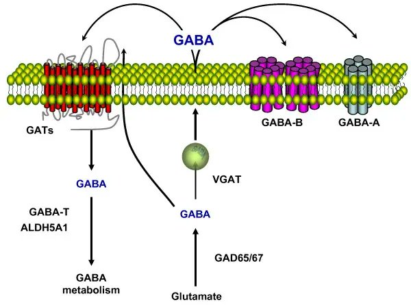
Newly synthesized GABA is packaged into synaptic vesicles by the vesicular GABA transporter (VGAT / SLC32A1), which uses a proton gradient.
Upon arrival of an action potential and calcium influx, vesicles undergo exocytosis, releasing GABA into the synaptic cleft.
Clearance occurs rapidly to terminate signaling and recycle the transmitter.
This is mediated by high-affinity, sodium- and chloride-dependent GABA transporters.
The predominant transporter is GAT1 (SLC6A1), expressed on both presynaptic GABAergic terminals and astrocytes. GAT1 accounts for ~70–85% of GABA reuptake.
A smaller contribution comes from GAT3 (SLC6A11) on astrocytes.
13
Once inside the cell, GABA is primarily metabolized through the GABA shunt, a bypass of the tricarboxylic acid (TCA) cycle that tightly links inhibitory neurotransmission to energy metabolism:
GABA transaminase (GABA-T / ABAT), a mitochondrial enzyme, transaminates GABA with α-ketoglutarate to produce succinic semialdehyde and glutamate (which can be reused for GABA synthesis). This step also requires P5P as a cofactor.
Succinic semialdehyde dehydrogenase (SSADH / ALDH5A1) then oxidizes succinic semialdehyde to succinate, generating NADH in the process.
Succinate enters the TCA cycle, supporting ATP production.
The net result of the GABA shunt is the conversion of α-ketoglutarate to succinate while recycling glutamate and contributing ~10–20% of neuronal energy under basal conditions (more during high activity).
This pathway links inhibitory neurotransmission directly to cellular energy metabolism.
So GABA counterbalances glutamate’s excitatory signals.
By opening chloride channels (primarily via GABAA receptors), it produces rapid hyperpolarization, or shunting inhibition, that decreases the probability of neuronal firing and helps maintain balanced brain activity.
Now GABAergic interneurons, including parvalbumin-, somatostatin-, and chandelier-positive cells, provide precise temporal and spatial control over pyramidal neuron firing.
They sculpt cortical rhythms (such as gamma oscillations), sharpen sensory processing, and support higher-order cognition.
It’s totally normal at this point to think “what the fuck does that even mean” so here’s an analogy we’ve previously used.
Imagine that your brain is a huge classroom full of kids (that represent neurons) who all want to talk at once.
GABAergic interneurons are like the teachers who keep everything under control.
There are three special teachers:
Parvalbumin teachers are the fastest and loudest teachers, telling everyone to be quiet.
Somatostatin teachers are the more patient ones, walking around and gently calming down the kids who are getting too excited on their own desks (that represent dendrites).
Chandelier teachers decide exactly when a kid is allowed to speak.
Now when it comes to being medically/biochemically accurate:
Parvalbumin-positive (PV+) interneurons are fast-spiking cells that provide powerful perisomatic inhibition, wrapping around the cell body and proximal dendrites of pyramidal neurons.
Their rapid kinetics enable them to synchronize large populations of pyramidal cells, generating high-frequency gamma oscillations (30–80 Hz).
These oscillations are critical for attention, working memory, sensory binding, and conscious perception.
PV+ interneurons are particularly vulnerable to oxidative stress and are frequently impaired in schizophrenia, autism spectrum disorder, and epilepsy.
Somatostatin-positive (SST+) interneurons primarily target distal dendrites of pyramidal neurons.
By modulating dendritic calcium spikes and excitatory input integration, they regulate synaptic plasticity, dendritic computation, and the gating of information flow.
SST+ cells are key players in feedback inhibition and are involved in shaping contextual and emotional processing in the hippocampus and cortex.
Chandelier cells, a specialized subtype of GABAergic interneuron, form distinctive “cartridge” synapses exclusively onto the axon initial segment of pyramidal neurons, the exact site where action potentials are generated.
This strategic positioning allows chandelier cells to exert powerful, high-fidelity control over pyramidal neuron output, acting as a final “gatekeeper” for neuronal firing.
Their dysfunction, whether through reduced numbers, impaired function, or altered synaptic strength, is a common feature in conditions characterized by E/I imbalance, including schizophrenia, autism, epilepsy, and major depressive disorder.
GABA transaminase (GABA-T / ABAT), a mitochondrial enzyme, transaminates GABA with α-ketoglutarate to produce succinic semialdehyde and glutamate (which can be reused for GABA synthesis). This step also requires P5P as a cofactor.
Succinic semialdehyde dehydrogenase (SSADH / ALDH5A1) then oxidizes succinic semialdehyde to succinate, generating NADH in the process.
Succinate enters the TCA cycle, supporting ATP production.
The net result of the GABA shunt is the conversion of α-ketoglutarate to succinate while recycling glutamate and contributing ~10–20% of neuronal energy under basal conditions (more during high activity).
This pathway links inhibitory neurotransmission directly to cellular energy metabolism.
So GABA counterbalances glutamate’s excitatory signals.
By opening chloride channels (primarily via GABAA receptors), it produces rapid hyperpolarization, or shunting inhibition, that decreases the probability of neuronal firing and helps maintain balanced brain activity.
Now GABAergic interneurons, including parvalbumin-, somatostatin-, and chandelier-positive cells, provide precise temporal and spatial control over pyramidal neuron firing.
They sculpt cortical rhythms (such as gamma oscillations), sharpen sensory processing, and support higher-order cognition.
It’s totally normal at this point to think “what the fuck does that even mean” so here’s an analogy we’ve previously used.
Imagine that your brain is a huge classroom full of kids (that represent neurons) who all want to talk at once.
GABAergic interneurons are like the teachers who keep everything under control.
There are three special teachers:
Parvalbumin teachers are the fastest and loudest teachers, telling everyone to be quiet.
Somatostatin teachers are the more patient ones, walking around and gently calming down the kids who are getting too excited on their own desks (that represent dendrites).
Chandelier teachers decide exactly when a kid is allowed to speak.
Now when it comes to being medically/biochemically accurate:
Parvalbumin-positive (PV+) interneurons are fast-spiking cells that provide powerful perisomatic inhibition, wrapping around the cell body and proximal dendrites of pyramidal neurons.
Their rapid kinetics enable them to synchronize large populations of pyramidal cells, generating high-frequency gamma oscillations (30–80 Hz).
These oscillations are critical for attention, working memory, sensory binding, and conscious perception.
PV+ interneurons are particularly vulnerable to oxidative stress and are frequently impaired in schizophrenia, autism spectrum disorder, and epilepsy.
Somatostatin-positive (SST+) interneurons primarily target distal dendrites of pyramidal neurons.
By modulating dendritic calcium spikes and excitatory input integration, they regulate synaptic plasticity, dendritic computation, and the gating of information flow.
SST+ cells are key players in feedback inhibition and are involved in shaping contextual and emotional processing in the hippocampus and cortex.
Chandelier cells, a specialized subtype of GABAergic interneuron, form distinctive “cartridge” synapses exclusively onto the axon initial segment of pyramidal neurons, the exact site where action potentials are generated.
This strategic positioning allows chandelier cells to exert powerful, high-fidelity control over pyramidal neuron output, acting as a final “gatekeeper” for neuronal firing.
Their dysfunction, whether through reduced numbers, impaired function, or altered synaptic strength, is a common feature in conditions characterized by E/I imbalance, including schizophrenia, autism, epilepsy, and major depressive disorder.
14
Now just like with all neurotransmitters, GABA signaling extends well beyond the central nervous system.
For example, in the pancreas, β-cells synthesize and co-release GABA along with insulin.
Once released, GABA acts on neighboring α-cells to inhibit glucagon secretion and exerts autocrine effects that promote β-cell survival and proliferation.
Or in the gastrointestinal tract, enteric neurons and certain gut bacteria, particularly strains of Lactobacillus and Bifidobacterium (Lactobacillus brevis, Bifidobacterium dentium, and others) synthesize GABA via their own GAD homologues which influences gut motility, secretion, and communication along the gut-brain axis via vagal and systemic pathways.
Also dysbiosis is associated with lower GABA levels and increased anxiety-like behavior in animal models and probiotic interventions sometimes raise brain GABA.
Additionally, many immune cells (including T cells and macrophages) express functional GABAA and GABAB receptors, through which GABA exerts anti-inflammatory and immunoregulatory effects by suppressing pro-inflammatory cytokines and promoting regulatory immune responses.
For example, in the pancreas, β-cells synthesize and co-release GABA along with insulin.
Once released, GABA acts on neighboring α-cells to inhibit glucagon secretion and exerts autocrine effects that promote β-cell survival and proliferation.
Or in the gastrointestinal tract, enteric neurons and certain gut bacteria, particularly strains of Lactobacillus and Bifidobacterium (Lactobacillus brevis, Bifidobacterium dentium, and others) synthesize GABA via their own GAD homologues which influences gut motility, secretion, and communication along the gut-brain axis via vagal and systemic pathways.
Also dysbiosis is associated with lower GABA levels and increased anxiety-like behavior in animal models and probiotic interventions sometimes raise brain GABA.
Additionally, many immune cells (including T cells and macrophages) express functional GABAA and GABAB receptors, through which GABA exerts anti-inflammatory and immunoregulatory effects by suppressing pro-inflammatory cytokines and promoting regulatory immune responses.
15
Now unfortunately, there are no routine clinical blood tests for brain GABA levels (plasma GABA correlates poorly with central concentrations).
Assessment therefore relies on:
Detailed symptom pattern recognition.
Response to empirical GABA-supportive interventions (magnesium, P5P, l-theanine and everything that we’ll discuss later).
Advanced imaging (MRS, PET) or EEG if for some reason are available, typically in research or specialty settings.
Exclusion of mimics/ruling out conditions that can produce overlapping symptoms, including thyroid dysfunction, vitamin B12 or folate deficiency, sleep apnea, and substance use or withdrawal.
Genetic testing for variants in GAD1/GAD2, SLC6A1 (GAT1), GABR subunit genes, or KCC2/NKCC1 can alsp be informative.
Assessment therefore relies on:
Detailed symptom pattern recognition.
Response to empirical GABA-supportive interventions (magnesium, P5P, l-theanine and everything that we’ll discuss later).
Advanced imaging (MRS, PET) or EEG if for some reason are available, typically in research or specialty settings.
Exclusion of mimics/ruling out conditions that can produce overlapping symptoms, including thyroid dysfunction, vitamin B12 or folate deficiency, sleep apnea, and substance use or withdrawal.
Genetic testing for variants in GAD1/GAD2, SLC6A1 (GAT1), GABR subunit genes, or KCC2/NKCC1 can alsp be informative.
16
But in general, modern lifestyles challenge GABAergic function through multiple converging mechanisms such as:
Chronic stress/glucocorticoid exposure (which downregulates GAD and GABAA receptor expression)
Micronutrient deficiencies
Neuroinflammation, mitochondrial impairment
Sleep disruption/harmed circadian health
Repeated exposure to GABAergic modulators such as alcohol that produce tolerance and rebound hyperexcitability.
Mitochondrial dysfunction
Hormonal imbalances
For example, GABA is not made from scratch, it is synthesized directly from glutamate via GAD as we saw in the previous article.
But the precursor pool of glutamate is generated inside the mitochondria.
So if mitochondria are damaged, they cannot export enough glutamate to the cytosol and without the raw building blocks, neurons cannot synthesize adequate amounts of GABA.
In general, GABA synthesis (GAD), vesicular loading (VGAT), reuptake (GAT1, Na⁺/K⁺-ATPase dependent), and degradation (mitochondrial GABA-T and SSADH) are all energy-intensive.
Besides this, pro-inflammatory cytokines (IL-1β, TNF-α, IL-6) suppress GAD activity, impair GAT1 trafficking, promote GABAA receptor internalization, plus reactive oxygen and nitrogen species damage GABAergic interneurons, which are particularly vulnerable due to high metabolic demand and relatively low antioxidant capacity.
Chronic stress/glucocorticoid exposure (which downregulates GAD and GABAA receptor expression)
Micronutrient deficiencies
Neuroinflammation, mitochondrial impairment
Sleep disruption/harmed circadian health
Repeated exposure to GABAergic modulators such as alcohol that produce tolerance and rebound hyperexcitability.
Mitochondrial dysfunction
Hormonal imbalances
For example, GABA is not made from scratch, it is synthesized directly from glutamate via GAD as we saw in the previous article.
But the precursor pool of glutamate is generated inside the mitochondria.
So if mitochondria are damaged, they cannot export enough glutamate to the cytosol and without the raw building blocks, neurons cannot synthesize adequate amounts of GABA.
In general, GABA synthesis (GAD), vesicular loading (VGAT), reuptake (GAT1, Na⁺/K⁺-ATPase dependent), and degradation (mitochondrial GABA-T and SSADH) are all energy-intensive.
Besides this, pro-inflammatory cytokines (IL-1β, TNF-α, IL-6) suppress GAD activity, impair GAT1 trafficking, promote GABAA receptor internalization, plus reactive oxygen and nitrogen species damage GABAergic interneurons, which are particularly vulnerable due to high metabolic demand and relatively low antioxidant capacity.
17
Then, in cases of hormonal issues such as low progesterone, high estrogen or hypothyroidism (underactive thyroid), the enzymatic activity required to synthesize GABA drops.
Thyroid hormones (T₃ and T₄) act as direct regulators of GAD activity.
Estrogen upregulates glutamate.
Your body converts progesterone into a neurosteroid called allopregnanolone (ALLO) that cts as a positive allosteric modulator for GABA-A.
This is partly why women on hormonal birth control are more likely to experience symptoms such as anxiety compared to non-users.
Then when it comes to micronutrient deficiencies the classic examples are B6, magnesium and zinc.
P5P is the essential cofactor for both GAD65 and GAD67.
So a deficiency impairs the glutamate-to-GABA conversion, rapidly lowering GABA synthesis.
Mg²⁺ acts as a positive allosteric modulator of GABAA receptors and supports GAD activity.
Hypomagnesemia increases neuronal excitability by reducing GABAergic inhibition and enhancing NMDA receptor function.
Zinc modulates GABAA receptor subtypes and transporter function.
Deficiencies in taurine, antioxidants, or B vitamins can of course further compromise GABA synthesis.
Taurine for example acts as a weak agonist and positive allosteric modulator of GABAA receptors.
It enhances GABA’s calming effects, especially at extrasynaptic receptors that mediate tonic inhibition.
Plus it has strong antioxidant and anti-inflammatory properties so it protects GABAergic interneurons (especially parvalbumin-positive cells) from oxidative stress and excitotoxicity.
So low taurine makes these vulnerable neurons more likely to become damaged or dysfunctional.
It also helps regulate intracellular chloride levels (via effects on NKCC1 and KCC2 transporters) which is important for keeping GABA inhibitory rather than excitatory and maintains cell volume and mitochondrial function in neurons and astrocytes.
Also, low taurine can lead to cellular stress that impairs GABA synthesis (GAD activity) and reuptake (GAT1 function).
Then, prolonged glucocorticoid exposure downregulates GAD67 expression, reduces GABAA receptor subunit mRNA (particularly α2 and γ2), and decreases parvalbumin interneuron density in the hippocampus and prefrontal cortex.
This creates a vicious cycle: low GABA → heightened stress reactivity → further HPA activation.
When it comes to things such as alcohol, its acute potentiation of GABAA receptors (especially δ-containing) might produce anxiolysis initially, but chronic exposure causes receptor downregulation, internalization, and tolerance.
Also obviously, chronic use of bezons leads to GABAA receptor desensitization, subunit switching (↓γ2, ↑α4), and dependence.
What’s also interesting is that heavy metals, pesticides (organophosphates, neonicotinoids), and certain environmental chemicals can also directly inhibit GAD or damage GABAergic neurons.
Thyroid hormones (T₃ and T₄) act as direct regulators of GAD activity.
Estrogen upregulates glutamate.
Your body converts progesterone into a neurosteroid called allopregnanolone (ALLO) that cts as a positive allosteric modulator for GABA-A.
This is partly why women on hormonal birth control are more likely to experience symptoms such as anxiety compared to non-users.
Then when it comes to micronutrient deficiencies the classic examples are B6, magnesium and zinc.
P5P is the essential cofactor for both GAD65 and GAD67.
So a deficiency impairs the glutamate-to-GABA conversion, rapidly lowering GABA synthesis.
Mg²⁺ acts as a positive allosteric modulator of GABAA receptors and supports GAD activity.
Hypomagnesemia increases neuronal excitability by reducing GABAergic inhibition and enhancing NMDA receptor function.
Zinc modulates GABAA receptor subtypes and transporter function.
Deficiencies in taurine, antioxidants, or B vitamins can of course further compromise GABA synthesis.
Taurine for example acts as a weak agonist and positive allosteric modulator of GABAA receptors.
It enhances GABA’s calming effects, especially at extrasynaptic receptors that mediate tonic inhibition.
Plus it has strong antioxidant and anti-inflammatory properties so it protects GABAergic interneurons (especially parvalbumin-positive cells) from oxidative stress and excitotoxicity.
So low taurine makes these vulnerable neurons more likely to become damaged or dysfunctional.
It also helps regulate intracellular chloride levels (via effects on NKCC1 and KCC2 transporters) which is important for keeping GABA inhibitory rather than excitatory and maintains cell volume and mitochondrial function in neurons and astrocytes.
Also, low taurine can lead to cellular stress that impairs GABA synthesis (GAD activity) and reuptake (GAT1 function).
Then, prolonged glucocorticoid exposure downregulates GAD67 expression, reduces GABAA receptor subunit mRNA (particularly α2 and γ2), and decreases parvalbumin interneuron density in the hippocampus and prefrontal cortex.
This creates a vicious cycle: low GABA → heightened stress reactivity → further HPA activation.
When it comes to things such as alcohol, its acute potentiation of GABAA receptors (especially δ-containing) might produce anxiolysis initially, but chronic exposure causes receptor downregulation, internalization, and tolerance.
Also obviously, chronic use of bezons leads to GABAA receptor desensitization, subunit switching (↓γ2, ↑α4), and dependence.
What’s also interesting is that heavy metals, pesticides (organophosphates, neonicotinoids), and certain environmental chemicals can also directly inhibit GAD or damage GABAergic neurons.
18
After addressing these, other tools you can consider adding to support GABA include:
Selank
It’s a synthetic heptapeptide (tuftsin analog) that allosterically modulates GABA-A receptors, increases GABA gene expression, and stabilizes enkephalins (natural opioids) without direct benzodiazepine-site binding.
Lithium orotate
Lithium increases GABA levels in the brain, it upregulates GAD and some evidence suggests it increases GABA-B receptor sensitivity.
Cardio
It increases GAD67 expression via BDNF.
Mediation
Meditation, tai chi and breathwork seem to increase GAD expression
L-Theanine
Increases GABA and alpha waves
Taurine
It’s a GABAA agonist and antioxidant
Apigenin (from chamomile)
Binds benzodiazepine site on GABAA
Lemon Balm
Inhibits GABA-T (reduces breakdown)
Curcumin
It’s an anti-inflammatory that seems to protect and upregulate GAD
Ketosis
It provides more ketone bodies (especially β-hydroxybutyrate), which directly upregulate GAD.
It also supplies more acetyl-CoA, which supports the TCA cycle, provides precursors for GABA production and even increases production of allopregnanolone and other neurosteroids that act as powerful positive allosteric modulators of GABA-A receptors (especially δ-containing ones for tonic inhibition).
Selank
It’s a synthetic heptapeptide (tuftsin analog) that allosterically modulates GABA-A receptors, increases GABA gene expression, and stabilizes enkephalins (natural opioids) without direct benzodiazepine-site binding.
Lithium orotate
Lithium increases GABA levels in the brain, it upregulates GAD and some evidence suggests it increases GABA-B receptor sensitivity.
Cardio
It increases GAD67 expression via BDNF.
Mediation
Meditation, tai chi and breathwork seem to increase GAD expression
L-Theanine
Increases GABA and alpha waves
Taurine
It’s a GABAA agonist and antioxidant
Apigenin (from chamomile)
Binds benzodiazepine site on GABAA
Lemon Balm
Inhibits GABA-T (reduces breakdown)
Curcumin
It’s an anti-inflammatory that seems to protect and upregulate GAD
Ketosis
It provides more ketone bodies (especially β-hydroxybutyrate), which directly upregulate GAD.
It also supplies more acetyl-CoA, which supports the TCA cycle, provides precursors for GABA production and even increases production of allopregnanolone and other neurosteroids that act as powerful positive allosteric modulators of GABA-A receptors (especially δ-containing ones for tonic inhibition).
19
That's all.
Now if you are interested in brain health overall, i will soon update this: fitandball.gumroad.com/l/brain20?layo…
Now if you are interested in brain health overall, i will soon update this: fitandball.gumroad.com/l/brain20?layo…
20
And if you liked this thread and found it useful, make sure to leave a like/RT.
George Ferman@Helios_Movement
How to start getting out of depression, ADHD, OCD, anxiety and panic disorders by utilizing GABA.
Thread🧵
Thread🧵
