If you give me 17 minutes, i will help you battle depression more...

George Ferman@Helios_Movement
125 views
Nov 29, 2025
~22 min read
Advertisement
1
If you give me 17 minutes, i will help you battle depression more effectively than everything you've tried so far.
This is the most in-depth guide about depression, discussing:
-Common misunderstandings when it comes to depression
-The genetic influences
-General tests you can take
-The hormonal influences
-Why where you live is very important
-Something more effective and safer than SSRIs
-Why, no matter how much you work on your mental patterns, you're still depressed
-Why you might be thinking about depression the wrong way
and much more.
Thread🧵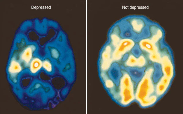
This is the most in-depth guide about depression, discussing:
-Common misunderstandings when it comes to depression
-The genetic influences
-General tests you can take
-The hormonal influences
-Why where you live is very important
-Something more effective and safer than SSRIs
-Why, no matter how much you work on your mental patterns, you're still depressed
-Why you might be thinking about depression the wrong way
and much more.
Thread🧵
2
*Standard disclaimer that nothing in this thread should be used as a substitute for medical advice*
It's George.
Let's start this thread with the following: there's no "one thing" that causes depression.
Depression is driven by a complex interplay of lifestyle factors, genetics, hormones and much more.
Now in order for someone to meet the diagnostic criteria for unipolar depression (major depressive disorder (MDD)), he must have at least 5 of the following symptoms that also cause him significant distress (impairment in social, occupational and other areas of his life)
The symptoms include:
- Anhedonia: a diminished ability to experience pleasure, interest or motivation in previously enjoyable activities for an extended time period.
-Significant weight loss or gain (more than 5% of body weight in a month).
-Insomnia or hypersomnia.
-Psychomotor changes such as agitation.
-Severe fatigue.
-Excessive or inappropriate guilt or feelings of inadequacy.
-Serious trouble focusing, making decisions or remembering.
-Recurrent thoughts of death, suicidal ideation or suicide attempts.
So if you don't have at least 5 of these symptoms, you might be mislabeling what you are feeling as depression.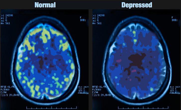
It's George.
Let's start this thread with the following: there's no "one thing" that causes depression.
Depression is driven by a complex interplay of lifestyle factors, genetics, hormones and much more.
Now in order for someone to meet the diagnostic criteria for unipolar depression (major depressive disorder (MDD)), he must have at least 5 of the following symptoms that also cause him significant distress (impairment in social, occupational and other areas of his life)
The symptoms include:
- Anhedonia: a diminished ability to experience pleasure, interest or motivation in previously enjoyable activities for an extended time period.
-Significant weight loss or gain (more than 5% of body weight in a month).
-Insomnia or hypersomnia.
-Psychomotor changes such as agitation.
-Severe fatigue.
-Excessive or inappropriate guilt or feelings of inadequacy.
-Serious trouble focusing, making decisions or remembering.
-Recurrent thoughts of death, suicidal ideation or suicide attempts.
So if you don't have at least 5 of these symptoms, you might be mislabeling what you are feeling as depression.
3
Now if these criteria were not enough to convince someone that when a person is depressed, his body literally changes, hypofrontality, particularly in the dorsolateral prefrontal cortex (DLPFC), that is well-documented in depression.
Hypofrontality basically = mitochondrial + vascular + synaptic failure.
This is partly why depressed people have:
-Impaired executive function/difficulty with decision-making
-Have emotional dysregulation and shut down, going into anhedonia
This is why, in this thread we will be focusing on:
-Mitochondrial health
- Cardiovascular health
-Gut health
-Things such as BDNF, GDNF, NGF etc
in certain parts of it.
Just a single cortical neuron utilizes approximately 4.7 billion ATPs per second in a resting human brain for example.
So if you are depressed, you should acknowledge the impact that your body can have and accept that it's not "all in your head".
You can go through thousands of hours of psychotherapy, read every book you can find, change every life circumstance you can think of, but if your body is not healthy at a sufficient level, your depression will still be there.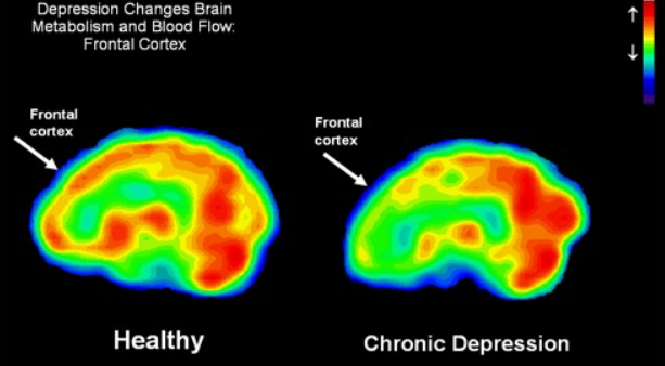
Hypofrontality basically = mitochondrial + vascular + synaptic failure.
This is partly why depressed people have:
-Impaired executive function/difficulty with decision-making
-Have emotional dysregulation and shut down, going into anhedonia
This is why, in this thread we will be focusing on:
-Mitochondrial health
- Cardiovascular health
-Gut health
-Things such as BDNF, GDNF, NGF etc
in certain parts of it.
Just a single cortical neuron utilizes approximately 4.7 billion ATPs per second in a resting human brain for example.
So if you are depressed, you should acknowledge the impact that your body can have and accept that it's not "all in your head".
You can go through thousands of hours of psychotherapy, read every book you can find, change every life circumstance you can think of, but if your body is not healthy at a sufficient level, your depression will still be there.
4
And finally, if these weren't enough, depression has been described as a neuroimmune disorder as well.
An example of this is the Reward and Immune Systems in Emotion (RISE) study, where researchers focused on the interplay between the brain’s reward system and systemic inflammation when it comes to depression.
Here's some background on it: the RISE study was a three-year longitudinal investigation that was tracking approximately 300 adolescents aged 13–16 from the Philadelphia area, all without a prior diagnosis of MDD.
This age range was strategic, as epidemiological data show a sharp rise in MDD incidence between ages 15–18, coinciding with peaks in reward system development and the emergence of pro-inflammatory phenotypes during adolescence.
The study’s longitudinal design spans three years, with comprehensive assessments at Time 1 (T1), Time 3 (T3), and Time 5 (T5) annually, and interim evaluations at Time 2 (T2) and Time 4 (T4) every six months.
By following teens over time, RISE aimed to capture dynamic changes in reward processing and inflammation that may precede depressive symptoms.
Participants were selected based on their self-reported reward responsiveness (RR), measured using the Behavioral Activation System (BAS) subscale of the Behavioral Inhibition System/Behavioral Activation System (BIS/BAS) Scales.
The study oversampled teens with low RR (0–20th percentile) to increase the likelihood of observing MDD onsets, as low RR is a hypothesized vulnerability for depression.
The comprehensive methodology included:
-Blood Draws: Fasting blood samples at T1, T3, and T5 quantify inflammatory biomarkers like C-reactive protein (CRP), interleukin-6 (IL-6), IL-8, IL-10, and tumor necrosis factor-alpha (TNF-α) using a high-sensitivity microfluidic platform.
-Neuroimaging: Functional magnetic resonance imaging (fMRI) at T1, T3, and T5 measures neural activity and connectivity in reward-related brain regions, such as the ventral striatum (VS) and orbitofrontal cortex (OFC), during tasks like the Monetary Incentive Delay (MID) Task and the Chatroom Interact Task.
These tasks assess responses to monetary rewards such as winning $5 and social rewards.
-Behavioral and Self-Report Measures: Tasks like the Card Arranging Reward Responsivity Objective Test (CARROT) and Delay Discounting Task evaluate behavioral RR, while questionnaires like the Sensitivity to Punishment/Sensitivity to Reward Questionnaire (SPSRQ) and Positive Valence Systems Scale (PVSS) capture subjective reward sensitivity.
-Life Events and Adversity: The Life Events Scale (LES) and Life Events Interview (LEI) track reward-relevant stressors such as goal failures and stress generation, while the Childhood Life Events Scale (CLES) assesses early adversity. Socioeconomic status (SES) and pubertal development (Pubertal Development Scale) were also evaluated as potential moderators.
-Inflammation-Enhancing Behaviors: Measures like the Adolescent Alcohol and Drug Involvement Scale (AADIS), Dietary Screening Questionnaire (DSQ), Pittsburgh Sleep Quality Index (PSQI), and actigraphy assess behaviors like substance use, poor diet, and sleep disruption that may amplify inflammation.
Now here's what these showed:
(1) low RR or its stunted development will correlate with elevated inflammation
(2) both high inflammation and low RR will independently predict depressive symptoms and MDD onset
(3) their interaction will amplify this risk
and
(4) early adversity will moderate, and behaviors like poor diet or sleep disruption will mediate, these associations.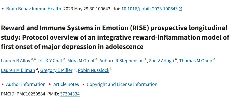
An example of this is the Reward and Immune Systems in Emotion (RISE) study, where researchers focused on the interplay between the brain’s reward system and systemic inflammation when it comes to depression.
Here's some background on it: the RISE study was a three-year longitudinal investigation that was tracking approximately 300 adolescents aged 13–16 from the Philadelphia area, all without a prior diagnosis of MDD.
This age range was strategic, as epidemiological data show a sharp rise in MDD incidence between ages 15–18, coinciding with peaks in reward system development and the emergence of pro-inflammatory phenotypes during adolescence.
The study’s longitudinal design spans three years, with comprehensive assessments at Time 1 (T1), Time 3 (T3), and Time 5 (T5) annually, and interim evaluations at Time 2 (T2) and Time 4 (T4) every six months.
By following teens over time, RISE aimed to capture dynamic changes in reward processing and inflammation that may precede depressive symptoms.
Participants were selected based on their self-reported reward responsiveness (RR), measured using the Behavioral Activation System (BAS) subscale of the Behavioral Inhibition System/Behavioral Activation System (BIS/BAS) Scales.
The study oversampled teens with low RR (0–20th percentile) to increase the likelihood of observing MDD onsets, as low RR is a hypothesized vulnerability for depression.
The comprehensive methodology included:
-Blood Draws: Fasting blood samples at T1, T3, and T5 quantify inflammatory biomarkers like C-reactive protein (CRP), interleukin-6 (IL-6), IL-8, IL-10, and tumor necrosis factor-alpha (TNF-α) using a high-sensitivity microfluidic platform.
-Neuroimaging: Functional magnetic resonance imaging (fMRI) at T1, T3, and T5 measures neural activity and connectivity in reward-related brain regions, such as the ventral striatum (VS) and orbitofrontal cortex (OFC), during tasks like the Monetary Incentive Delay (MID) Task and the Chatroom Interact Task.
These tasks assess responses to monetary rewards such as winning $5 and social rewards.
-Behavioral and Self-Report Measures: Tasks like the Card Arranging Reward Responsivity Objective Test (CARROT) and Delay Discounting Task evaluate behavioral RR, while questionnaires like the Sensitivity to Punishment/Sensitivity to Reward Questionnaire (SPSRQ) and Positive Valence Systems Scale (PVSS) capture subjective reward sensitivity.
-Life Events and Adversity: The Life Events Scale (LES) and Life Events Interview (LEI) track reward-relevant stressors such as goal failures and stress generation, while the Childhood Life Events Scale (CLES) assesses early adversity. Socioeconomic status (SES) and pubertal development (Pubertal Development Scale) were also evaluated as potential moderators.
-Inflammation-Enhancing Behaviors: Measures like the Adolescent Alcohol and Drug Involvement Scale (AADIS), Dietary Screening Questionnaire (DSQ), Pittsburgh Sleep Quality Index (PSQI), and actigraphy assess behaviors like substance use, poor diet, and sleep disruption that may amplify inflammation.
Now here's what these showed:
(1) low RR or its stunted development will correlate with elevated inflammation
(2) both high inflammation and low RR will independently predict depressive symptoms and MDD onset
(3) their interaction will amplify this risk
and
(4) early adversity will moderate, and behaviors like poor diet or sleep disruption will mediate, these associations.
5
Now the brain’s reward system, centered in the cortico-striatal circuit governs our ability to anticipate and enjoy rewards.
When depressed, this system is often blunted, leading to anhedonia (a hallmark symptom of people who individuals struggle to find pleasure in activities).
Now inflammation, the body’s response to injury or infection, involves immune cells releasing cytokines (such as IL-6 and/or TNF-α) and acute-phase proteins like CRP.
Chronic, low-grade inflammation is implicated in depression, with meta-analyses showing elevated IL-6, TNF-α, and CRP in MDD patients compared to healthy controls.
Peripheral cytokines can access the brain via leaky regions of the blood-brain barrier (BBB), active transport, or vagal nerve signaling, where they disrupt dopamine synthesis and reward processing.
For instance, studies show that inflammatory stimuli (such as interferon-α treatment for hepatitis C) reduce VS activation to rewards, mimicking anhedonia.
The sources of inflammation in depression are diverse and include:
-Stress and trauma: Psychosocial stressors, especially early life adversity (abuse, neglect etc), activate the hypothalamic-pituitary-adrenal (HPA) axis, increasing cortisol and pro-inflammatory cytokines like IL-6 and TNF-α. The Dunedin study for example found that childhood maltreatment doubles the risk of chronic inflammation, which persists into adulthood and heightens depression risk.
-Diet: Western diets high in saturated fats and sugars increase inflammation, while Mediterranean diets rich in fiber, omega-3s, and phytochemicals reduce it. For example, the Nurses’ Health Study showed that “prudent” diets lower CRP and IL-6, while “Western” diets elevate them.
-Physical Inactivity: Sedentary behavior is linked to higher CRP and IL-6.
-Obesity: Adipose tissue produces cytokines like IL-6 and TNF-α, contributing to systemic inflammation. A meta-analysis found a bidirectional link, with obesity increasing depression risk by 55% and depression increasing obesity risk by 58%.
-Smoking: Cigarette smoke contains free radicals and metals that trigger oxidative stress and inflammation, elevating CRP, IL-6, and TNF-α. Depressed smokers show higher inflammation than non-depressed smokers.
-Gut Permeability: Increased gut permeability (“leaky gut”) allows bacterial lipopolysaccharides (LPS) to enter the bloodstream, triggering immune responses via Toll-like receptor-4 (TLR4).Studies show elevated IgA/IgM against gram-negative bacteria in MDD, suggesting bacterial translocation drives inflammation.
-Sleep Disruption: Poor sleep increases IL-6 and TNF-α, potentially via sympathetic nervous system activation. Plenty of meta-analysis by now have confirmed that insomnia is a risk factor for depression.
-Vitamin D Deficiency: Low vitamin D levels, common in Western populations, are linked to higher TNF-α and IL-6, increasing depression risk.
Now the RISE study tests an integrative model where low RR and high inflammation form a bidirectional feedback loop.
Peripheral cytokines reduce dopamine signaling in the VS, blunting reward responsiveness and leading to anhedonia.
Other studies highlight additional mechanisms such as:
-The kynurenine pathway.
Cytokines activate indoleamine 2,3-dioxygenase (IDO), catabolizing tryptophan into kynurenine, reducing serotonin availability and contributing to depression.
-Or quinolinic acid, a metabolite of the kynurenine pathway has neurotoxic properties at excessive levels since it leads to:
NMDA overactivation.
Oxidative stress
Diverting tryptophan away from serotonin synthesis.
-BBB disruption.
Chronic stress or inflammation weakens the BBB, allowing peripheral immune cells (such as Th17 cells or monocytes) to infiltrate the brain, promoting neuroinflammation.

When depressed, this system is often blunted, leading to anhedonia (a hallmark symptom of people who individuals struggle to find pleasure in activities).
Now inflammation, the body’s response to injury or infection, involves immune cells releasing cytokines (such as IL-6 and/or TNF-α) and acute-phase proteins like CRP.
Chronic, low-grade inflammation is implicated in depression, with meta-analyses showing elevated IL-6, TNF-α, and CRP in MDD patients compared to healthy controls.
Peripheral cytokines can access the brain via leaky regions of the blood-brain barrier (BBB), active transport, or vagal nerve signaling, where they disrupt dopamine synthesis and reward processing.
For instance, studies show that inflammatory stimuli (such as interferon-α treatment for hepatitis C) reduce VS activation to rewards, mimicking anhedonia.
The sources of inflammation in depression are diverse and include:
-Stress and trauma: Psychosocial stressors, especially early life adversity (abuse, neglect etc), activate the hypothalamic-pituitary-adrenal (HPA) axis, increasing cortisol and pro-inflammatory cytokines like IL-6 and TNF-α. The Dunedin study for example found that childhood maltreatment doubles the risk of chronic inflammation, which persists into adulthood and heightens depression risk.
-Diet: Western diets high in saturated fats and sugars increase inflammation, while Mediterranean diets rich in fiber, omega-3s, and phytochemicals reduce it. For example, the Nurses’ Health Study showed that “prudent” diets lower CRP and IL-6, while “Western” diets elevate them.
-Physical Inactivity: Sedentary behavior is linked to higher CRP and IL-6.
-Obesity: Adipose tissue produces cytokines like IL-6 and TNF-α, contributing to systemic inflammation. A meta-analysis found a bidirectional link, with obesity increasing depression risk by 55% and depression increasing obesity risk by 58%.
-Smoking: Cigarette smoke contains free radicals and metals that trigger oxidative stress and inflammation, elevating CRP, IL-6, and TNF-α. Depressed smokers show higher inflammation than non-depressed smokers.
-Gut Permeability: Increased gut permeability (“leaky gut”) allows bacterial lipopolysaccharides (LPS) to enter the bloodstream, triggering immune responses via Toll-like receptor-4 (TLR4).Studies show elevated IgA/IgM against gram-negative bacteria in MDD, suggesting bacterial translocation drives inflammation.
-Sleep Disruption: Poor sleep increases IL-6 and TNF-α, potentially via sympathetic nervous system activation. Plenty of meta-analysis by now have confirmed that insomnia is a risk factor for depression.
-Vitamin D Deficiency: Low vitamin D levels, common in Western populations, are linked to higher TNF-α and IL-6, increasing depression risk.
Now the RISE study tests an integrative model where low RR and high inflammation form a bidirectional feedback loop.
Peripheral cytokines reduce dopamine signaling in the VS, blunting reward responsiveness and leading to anhedonia.
Other studies highlight additional mechanisms such as:
-The kynurenine pathway.
Cytokines activate indoleamine 2,3-dioxygenase (IDO), catabolizing tryptophan into kynurenine, reducing serotonin availability and contributing to depression.
-Or quinolinic acid, a metabolite of the kynurenine pathway has neurotoxic properties at excessive levels since it leads to:
NMDA overactivation.
Oxidative stress
Diverting tryptophan away from serotonin synthesis.
-BBB disruption.
Chronic stress or inflammation weakens the BBB, allowing peripheral immune cells (such as Th17 cells or monocytes) to infiltrate the brain, promoting neuroinflammation.
6
Now of course, some people might dismiss these and think that only SSRIs can help with depression.
The response to this argument is that:
-SSRIs have plenty of side effects
-Serotonin depletion does not cause depression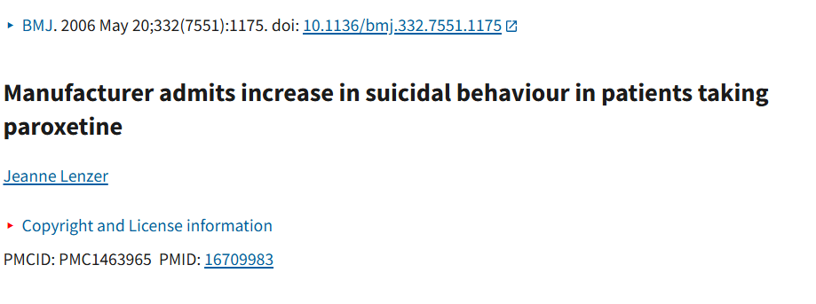

The response to this argument is that:
-SSRIs have plenty of side effects
-Serotonin depletion does not cause depression
7
A good idea is also to discuss some tests you can do in case you are struggling with depression.
Number 1: hsCRP.
This basically is a substance produced by the liver that increases in the presence of inflammation in the body and meta-analyses of >200 studies show elevated proinflammatory cytokines in ~30–40% of MDD patients.
Now, here's a list of common lifestyle factors and conditions that exacerbate systemic inflammation:
Being overweight increases everything (TNF-α, IL-6, IL-1β, MCP-1, IL-8 etc).
Trans fats that (mainly) increase TNF-α and IL-6. A high O6, low O3 ratio in the diet that (mainly) increases PGE2 and IL-8.
Anything that can lead to endotoxemia will (mainly) increase IL-6, TNF-α and IL-1β.
Alcohol (more than 1 shot a day) will increase IL-6, TNF-α and IL-1β. T2D/high blood sugar can elevate IL-6 and CRP.
Too much iron can increase IL-6 and CRP. Sleep deprivation increases IL-6, TNF-α, CRP.
Sleep apnea increases IL-6, IL-8, IL-1β.
Low muscle mass increases IL-6 and CRP. Sedentary behavior (>8 h/day) increases IL-6 and TNF-α.
A vitamin D deficiency increases IL-6, TNF-α and IL-17. Chronic stress also increases pretty much everything.
Mold mycotoxins increase IL-1β, IL-6 and TNF-α. Heavy metals IL-6, IL-1β and TNF-α (but less than mold).
A magnesium deficiency increases IL-6, CRP. A vitamin C deficiency increases IL-1β.
A zinc deficiency increases IL-6 and IL-1β.
A selenium deficiency increases TNF-α.
Overtraining increases IL-6 and IL-1β.
MCAS also increases pretty much everything. Melatonin suppression pretty much also increases almost everything down the line.
The following genetic variants:
Number 1: hsCRP.
This basically is a substance produced by the liver that increases in the presence of inflammation in the body and meta-analyses of >200 studies show elevated proinflammatory cytokines in ~30–40% of MDD patients.
Now, here's a list of common lifestyle factors and conditions that exacerbate systemic inflammation:
Being overweight increases everything (TNF-α, IL-6, IL-1β, MCP-1, IL-8 etc).
Trans fats that (mainly) increase TNF-α and IL-6. A high O6, low O3 ratio in the diet that (mainly) increases PGE2 and IL-8.
Anything that can lead to endotoxemia will (mainly) increase IL-6, TNF-α and IL-1β.
Alcohol (more than 1 shot a day) will increase IL-6, TNF-α and IL-1β. T2D/high blood sugar can elevate IL-6 and CRP.
Too much iron can increase IL-6 and CRP. Sleep deprivation increases IL-6, TNF-α, CRP.
Sleep apnea increases IL-6, IL-8, IL-1β.
Low muscle mass increases IL-6 and CRP. Sedentary behavior (>8 h/day) increases IL-6 and TNF-α.
A vitamin D deficiency increases IL-6, TNF-α and IL-17. Chronic stress also increases pretty much everything.
Mold mycotoxins increase IL-1β, IL-6 and TNF-α. Heavy metals IL-6, IL-1β and TNF-α (but less than mold).
A magnesium deficiency increases IL-6, CRP. A vitamin C deficiency increases IL-1β.
A zinc deficiency increases IL-6 and IL-1β.
A selenium deficiency increases TNF-α.
Overtraining increases IL-6 and IL-1β.
MCAS also increases pretty much everything. Melatonin suppression pretty much also increases almost everything down the line.
The following genetic variants:
8
Number 2: 25(OH)D (vitamin D from the SUN).
Some studies have even found that 60 percent of the suicidal patients were deficient in Vitamin D.
And it's no wonder why. VDRs are expressed in most cell types and tissues throughout the body, including the immune system, brain, heart, pancreas, muscles, skin and endocrine glands.
So a "simple" vitamin D deficiency will create plenty of problems. BUT, remember that you need the sun (or a vitamin D lamp).
Some studies have even found that 60 percent of the suicidal patients were deficient in Vitamin D.
And it's no wonder why. VDRs are expressed in most cell types and tissues throughout the body, including the immune system, brain, heart, pancreas, muscles, skin and endocrine glands.
So a "simple" vitamin D deficiency will create plenty of problems. BUT, remember that you need the sun (or a vitamin D lamp).
9
Number 3: A thyroid panel (Thyroid Stimulating Hormone (TSH), Free Triiodothyroine (fT3), Free Serum Thyroxine (fT4) and Thyroid Antibodies), estradiol, free testosterone and total testosterone.
Any hormonal problem will have negative effects on our mental well being.
Testosterone for example can even promote the regeneration of myelin.
Any hormonal problem will have negative effects on our mental well being.
Testosterone for example can even promote the regeneration of myelin.
10
Number 4: LDL, trig:HDL ratio.
Even though it's unlikely to happen unless you are on statins or have some sort of liver issues, lower levels of LDL have been linked to a lot of mental health problems ranging from depression all the way to violence.
Even though it's unlikely to happen unless you are on statins or have some sort of liver issues, lower levels of LDL have been linked to a lot of mental health problems ranging from depression all the way to violence.
11
Number 5: BDNF report / BDNF–TrkB signaling.
A polymorphism such as the rs6265 is a common cause of "treatment resistant depression" and why approaches such as ketamine, esketamine and psilocybin have become so popular because these drugs bypass activity-dependent BDNF secretion.
Overall, lower serum/plasma BDNF and reduced hippocampal volume are among the most replicated findings in MDD.
A polymorphism such as the rs6265 is a common cause of "treatment resistant depression" and why approaches such as ketamine, esketamine and psilocybin have become so popular because these drugs bypass activity-dependent BDNF secretion.
Overall, lower serum/plasma BDNF and reduced hippocampal volume are among the most replicated findings in MDD.
12
Number 6: Anything related to HPA axis dysregulation (NR3C1, CRHR1, FKBP5, etc.)
Meta-analyses consistently show hyperactive HPA axis (elevated cortisol, impaired dexamethasone suppression test) in ~50% of MDD patients and genetic variants in NR3C1 (glucocorticoid receptor), CRHR1 (corticotropin-releasing hormone receptor 1), and FKBP5 are associated with MDD risk and worse antidepressant response.
If you have the FKBP5 rs1360780 T allele for example you have an exaggerated cortisol response and poorer SSRI response but better response to psychotherapy and ketamine/esketamine.
Meta-analyses consistently show hyperactive HPA axis (elevated cortisol, impaired dexamethasone suppression test) in ~50% of MDD patients and genetic variants in NR3C1 (glucocorticoid receptor), CRHR1 (corticotropin-releasing hormone receptor 1), and FKBP5 are associated with MDD risk and worse antidepressant response.
If you have the FKBP5 rs1360780 T allele for example you have an exaggerated cortisol response and poorer SSRI response but better response to psychotherapy and ketamine/esketamine.
13
Number 7: Anything related to circadian rhythm disruption (PER2, PER3, CLOCK, CRY1)
Clock gene polymorphisms (PER3, CLOCK 3111T/C) are associated with MDD.
Plus agomelatine (MT1/MT2 agonist + 5-HT2C antagonist) works largely via circadian resynchronization and is effective specifically in patients with sleep–circadian disturbances.
Example: PER3 VNTR 5/5 repeat genotype → extreme evening chronotype → 2–3× higher risk of depression and seasonal worsening.
Number 8: Elevated kynurenine/tryptophan ratio that is consistently found in MDD (and normalizes with successful treatment).
Ketamine and psilocybin for example both strongly inhibit IDO1 and shift the pathway back toward neuroprotection within hours.
Clock gene polymorphisms (PER3, CLOCK 3111T/C) are associated with MDD.
Plus agomelatine (MT1/MT2 agonist + 5-HT2C antagonist) works largely via circadian resynchronization and is effective specifically in patients with sleep–circadian disturbances.
Example: PER3 VNTR 5/5 repeat genotype → extreme evening chronotype → 2–3× higher risk of depression and seasonal worsening.
Number 8: Elevated kynurenine/tryptophan ratio that is consistently found in MDD (and normalizes with successful treatment).
Ketamine and psilocybin for example both strongly inhibit IDO1 and shift the pathway back toward neuroprotection within hours.
14
Number 9: MTHFR.
Mainly the 677TT genotype is associated with higher MDD risk and poorer SSRI response, but better response when l-methylfolate is added (supported by several RCTs).
Number 10: Urea breath test, stool antigen test for H. pylori antigens, breath tests that measure hydrogen, methane, or hydrogen sulfide after lactulose/glucose ingestion, a stool analysis for candida, fungal culture, CHROMagar Candida.
Number 11: Fasting Glucose, insulin and hemoglobin A1C.
Blood sugar and insulin are both associated with depression.
Number 12: Magnesium and zinc.
These two nutrients were also found very low in suicide victims.
Number 13: Lyme and Celiac's.
This might sound weird (and it should to be fair) but plenty of people who develop serious depression all of a sudden actually have Lyme (Dr. Robert Bransfield has spoken about this).
Also plenty of people have silent Celiac's and don't know it.
Mainly the 677TT genotype is associated with higher MDD risk and poorer SSRI response, but better response when l-methylfolate is added (supported by several RCTs).
Number 10: Urea breath test, stool antigen test for H. pylori antigens, breath tests that measure hydrogen, methane, or hydrogen sulfide after lactulose/glucose ingestion, a stool analysis for candida, fungal culture, CHROMagar Candida.
Number 11: Fasting Glucose, insulin and hemoglobin A1C.
Blood sugar and insulin are both associated with depression.
Number 12: Magnesium and zinc.
These two nutrients were also found very low in suicide victims.
Number 13: Lyme and Celiac's.
This might sound weird (and it should to be fair) but plenty of people who develop serious depression all of a sudden actually have Lyme (Dr. Robert Bransfield has spoken about this).
Also plenty of people have silent Celiac's and don't know it.
15
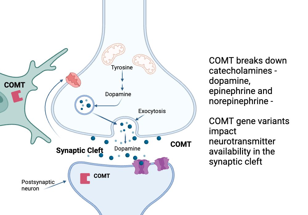
For more details on COMT and MTHFR, you can read this:

George Ferman@Helios_Movement
From anxiety, depression and insomnia all the way to the sad event of miscarriages, eczema and hair loss, a chronic health issue might be partly caused by SNPs in COMT and MTHFR that are getting quite popular these days.
But most advice regarding them is too complicated or too simplistic.
Here's the middle ground👇
Thread🧵
But most advice regarding them is too complicated or too simplistic.
Here's the middle ground👇
Thread🧵
16
Now let's move on to the rest of the suggestions.
Number 1: Work on your circadian rhythm (get bright natural light first thing in the day and block the artificial blue light as first steps).
To put in perspective how crucial this is, eating breakfast is linked to lower suic*de rates.
Plus: Spending 1.5 h/day in outdoor light is associated with a lower risk of depression, REGARDLESS of genetic risk, and a 1-hour daily of morning walk outside showed a 48% reduction in HDRS.
Now if you are still not convinced, just having light in your bedroom while you sleep makes you more depressed (tap in the pics).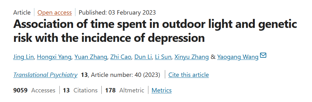


Number 1: Work on your circadian rhythm (get bright natural light first thing in the day and block the artificial blue light as first steps).
To put in perspective how crucial this is, eating breakfast is linked to lower suic*de rates.
Plus: Spending 1.5 h/day in outdoor light is associated with a lower risk of depression, REGARDLESS of genetic risk, and a 1-hour daily of morning walk outside showed a 48% reduction in HDRS.
Now if you are still not convinced, just having light in your bedroom while you sleep makes you more depressed (tap in the pics).
17
If you are still skeptical, chronotherapy has been one of the most effective and rapid therapies for treating depression.

18
Number 2: Start micronutrient maxing.
Your nutrition matters, a lot.
More than you probably want to (just tap in the pics).
For example:
-A meatless diet increases the frequency of depressive episodes.
-Thiamine and B12 decrease depressive episodes
-Suic*de victims often show low levels of zinc, magnesium and myo-inositol
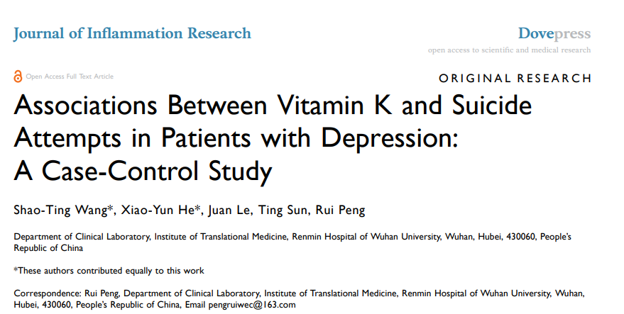
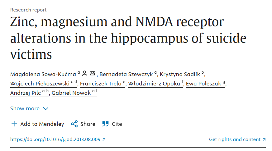
Your nutrition matters, a lot.
More than you probably want to (just tap in the pics).
For example:
-A meatless diet increases the frequency of depressive episodes.
-Thiamine and B12 decrease depressive episodes
-Suic*de victims often show low levels of zinc, magnesium and myo-inositol
19
Once again, you are probably underappreciating the impact that nutrients have on our well being.
Let's take B9 as an example.
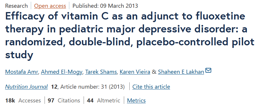
The study on the left found that prescription folic acid was associated with a 44% reduction in suicide attempts and self-harm in a large U.S. patient database of over 850,000 individuals.
Is this the BEST study? No.
Should you use folic acid instead of other forms? No.
Point being that low folate in general will:
-Harm the production of every single neurotransmitter (folate is required to make SAMe (S-adenosylmethionine) low folate → low SAMe → increased risk of depression, anxiety, bipolar disorder, schizophrenia and cognitive decline).
-Harm monoamine synthesis (via BH4 regeneration).
-Potentially cause neural tube defects (more and more doctors thankfully start testing for MTHFR SNPs on women who have had miscarriages).
-Harm the methylation cycle that's crucial for:
1. Gene expression.
2. Breaking down excess estrogen and catecholamines
3. Synthesis of melatonin, CoQ10, carnitine, creatine, phosphatidylcholine
and much more.
-Harm phase II detoxification.
-Increase homocysteine (helps convert homocysteine back to methionine).
-Harm cell turnover in the gut lining.
-Reduce T-cell production.
-Potentially cause anemia.
Etc.
And that's just one nutrient.
Let's take B9 as an example.
The study on the left found that prescription folic acid was associated with a 44% reduction in suicide attempts and self-harm in a large U.S. patient database of over 850,000 individuals.
Is this the BEST study? No.
Should you use folic acid instead of other forms? No.
Point being that low folate in general will:
-Harm the production of every single neurotransmitter (folate is required to make SAMe (S-adenosylmethionine) low folate → low SAMe → increased risk of depression, anxiety, bipolar disorder, schizophrenia and cognitive decline).
-Harm monoamine synthesis (via BH4 regeneration).
-Potentially cause neural tube defects (more and more doctors thankfully start testing for MTHFR SNPs on women who have had miscarriages).
-Harm the methylation cycle that's crucial for:
1. Gene expression.
2. Breaking down excess estrogen and catecholamines
3. Synthesis of melatonin, CoQ10, carnitine, creatine, phosphatidylcholine
and much more.
-Harm phase II detoxification.
-Increase homocysteine (helps convert homocysteine back to methionine).
-Harm cell turnover in the gut lining.
-Reduce T-cell production.
-Potentially cause anemia.
Etc.
And that's just one nutrient.
20
Number 3: Try to hang out with better people and at better places.
*Not a joke.
Number 4: Spend time in nature.
Walking in nature for example:
-Reduces amygdala activity (especially in women).
-It reduces cortisol quite fast (even 30 minutes spent in nature can lower cortisol by 30%-50%).
-Is one of the most effective ways to destimulate.
-Can increase levels of DHEA and adiponectin, which support our cardiovascular and overall metabolic health.

*Not a joke.
Number 4: Spend time in nature.
Walking in nature for example:
-Reduces amygdala activity (especially in women).
-It reduces cortisol quite fast (even 30 minutes spent in nature can lower cortisol by 30%-50%).
-Is one of the most effective ways to destimulate.
-Can increase levels of DHEA and adiponectin, which support our cardiovascular and overall metabolic health.
21
Number 5: Hit the IR sauna.
It's not only proven to be more effective than not only SSRIs but exercise as well, but it's also a great way to deal with the heavy metal load.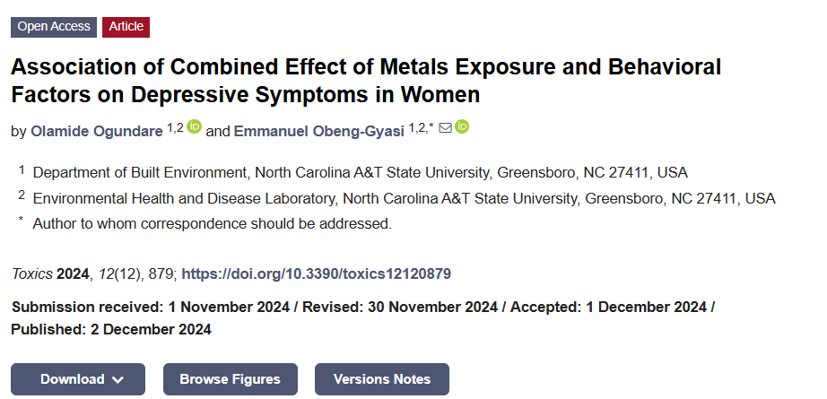

It's not only proven to be more effective than not only SSRIs but exercise as well, but it's also a great way to deal with the heavy metal load.
22
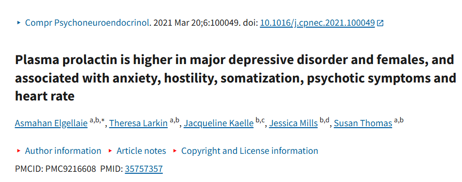
Number 6: Cut out the junk food and alcohol.
These are the fastest ways to lower some of the existing inflammation.
Just do it.
Number 7: Go test your hormones (a thyroid panel, insulin/IGF01, an androgen panel and an estrogen panel).
Point blank: A basic TRT dose might do more than 20000 SSRI pills for plenty of men.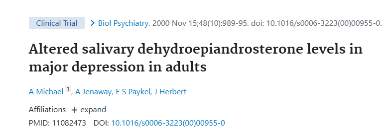
These are the fastest ways to lower some of the existing inflammation.
Just do it.
Number 7: Go test your hormones (a thyroid panel, insulin/IGF01, an androgen panel and an estrogen panel).
Point blank: A basic TRT dose might do more than 20000 SSRI pills for plenty of men.
23
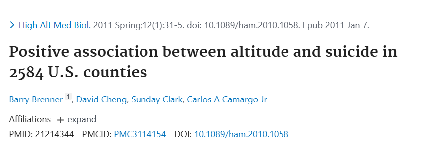
Number 8: Go to the beach.
I wish this was a joke but altitude is indeed one of the strongest geographic predictors of depression and su*cide rates.
When you increase the altitude, the reduced atmospheric pressure lowers blood oxygen, impairing mitochondrial ATP production in the brain (especially the prefrontal cortex).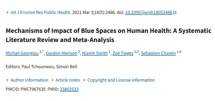

I wish this was a joke but altitude is indeed one of the strongest geographic predictors of depression and su*cide rates.
When you increase the altitude, the reduced atmospheric pressure lowers blood oxygen, impairing mitochondrial ATP production in the brain (especially the prefrontal cortex).
24
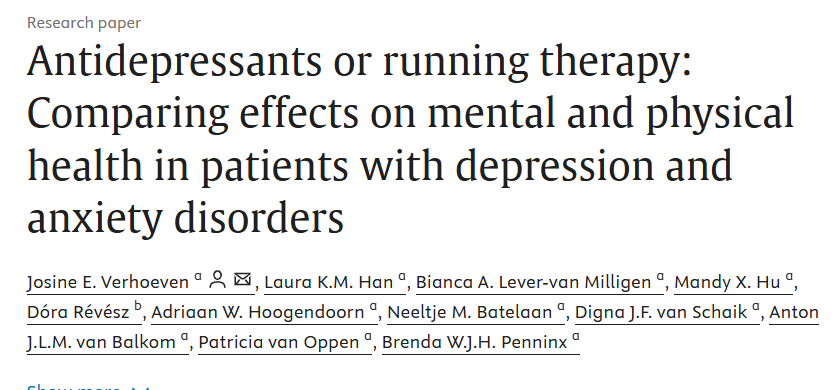
Number 9: Walk (like, a lot) and do your cardio daily.

25
Number 11: Work on your gut.
Our gut impacts our mood from the day we are born and i can make you develop OCD and even schizophrenia by changing its composition.


Our gut impacts our mood from the day we are born and i can make you develop OCD and even schizophrenia by changing its composition.
George Ferman@Helios_Movement
Most people will be too lazy to read this, but if you are struggling with gut issues such as:
-Bloating
-Constipation
-IBS
-Leaky gut
-SIBO
-SIFO
-Candida
Here's the ultimate plan for improving them.
Take 10 minutes to read this and you will be on the right track.
Thread🧵
-Bloating
-Constipation
-IBS
-Leaky gut
-SIBO
-SIFO
-Candida
Here's the ultimate plan for improving them.
Take 10 minutes to read this and you will be on the right track.
Thread🧵
26
Number 12: Research the following tools.
Tool 1: Red light therapy
-It boosts ATP production in neurons.
-It decreases pro-inflammatory cytokines (IL-1β, TNF-α), protecting the blood-brain barrier (BBB) and neurons,.
-It stimulates the release of brain-derived neurotrophic factor (BDNF).
-It increases cerebral blood flow, supporting nutrient delivery and waste removal.
-It reduces secondary brain damage in TBI and stroke by decreasing inflammation, excitotoxicity, and neuronal apoptosis.
Tool 2: Curcumin (boosts BDNF and NGF, reduces oxidative stress, inhibits pro-inflammatory pathways and upregulates antioxidant enzymes).
Tool 3: Royal jelly (mainly for the BBB).
Tool 4: Lion's mane (Increases NGF and BDNF like few things do).
Tool 5: Any form of magnesium besides oxide and citrate (enhances BDNF, supports synaptic plasticity, blocks glutamate excitotoxicity and promotes BBB stability)
Tool 6: NAC (boosts glutathione, reduces oxidative stress, supports BBB integrity and enhances BDNF)
Tool 7: ALCAR (supports BDNF, enhances mitochondrial function and it may protect BBB by reducing oxidative stress).
Tool 8: CoQ10 (Enhances mitochondrial function, reduces oxidative stress, supports BDNF/GDNF and protects the BBB).
Tool 9: Gingko biloba (enhances BDNF, improves cerebral blood flow and supports BBB integrity.)
Tool 10: Bacopa (Increases BDNF and NGF).
Tool 11: PQQ (enhances BDNF and NGF, supports mitochondrial biogenesis, and protects BBB)
Tool 12: Gotu kola (increases BDNF and NGF)
Tool 13: Phosphatidylserine (supports BDNF and stabilizes BBB).
Tool 14: Selank (enhances GABA receptor activity and increasing brain-derived neurotrophic factor).
Tool 15: Semax (boosts BDNF and NGF).
Tool 16: Cerebrolysin (mimics endogenous neurotrophic factors).
Tool 17: Huperzine A (enhances NGF and BDNF)
Tool 18: Saffron (boosts BDNF and NGF, reduces oxidative stress, and supports BBB)
Tool 19: Creatine (increases phosphocreatine stores in neurons, reduces oxidative stress, upregulates BDNF, supports endothelial cell stability in the BBB).
Tool 1: Red light therapy
-It boosts ATP production in neurons.
-It decreases pro-inflammatory cytokines (IL-1β, TNF-α), protecting the blood-brain barrier (BBB) and neurons,.
-It stimulates the release of brain-derived neurotrophic factor (BDNF).
-It increases cerebral blood flow, supporting nutrient delivery and waste removal.
-It reduces secondary brain damage in TBI and stroke by decreasing inflammation, excitotoxicity, and neuronal apoptosis.
Tool 2: Curcumin (boosts BDNF and NGF, reduces oxidative stress, inhibits pro-inflammatory pathways and upregulates antioxidant enzymes).
Tool 3: Royal jelly (mainly for the BBB).
Tool 4: Lion's mane (Increases NGF and BDNF like few things do).
Tool 5: Any form of magnesium besides oxide and citrate (enhances BDNF, supports synaptic plasticity, blocks glutamate excitotoxicity and promotes BBB stability)
Tool 6: NAC (boosts glutathione, reduces oxidative stress, supports BBB integrity and enhances BDNF)
Tool 7: ALCAR (supports BDNF, enhances mitochondrial function and it may protect BBB by reducing oxidative stress).
Tool 8: CoQ10 (Enhances mitochondrial function, reduces oxidative stress, supports BDNF/GDNF and protects the BBB).
Tool 9: Gingko biloba (enhances BDNF, improves cerebral blood flow and supports BBB integrity.)
Tool 10: Bacopa (Increases BDNF and NGF).
Tool 11: PQQ (enhances BDNF and NGF, supports mitochondrial biogenesis, and protects BBB)
Tool 12: Gotu kola (increases BDNF and NGF)
Tool 13: Phosphatidylserine (supports BDNF and stabilizes BBB).
Tool 14: Selank (enhances GABA receptor activity and increasing brain-derived neurotrophic factor).
Tool 15: Semax (boosts BDNF and NGF).
Tool 16: Cerebrolysin (mimics endogenous neurotrophic factors).
Tool 17: Huperzine A (enhances NGF and BDNF)
Tool 18: Saffron (boosts BDNF and NGF, reduces oxidative stress, and supports BBB)
Tool 19: Creatine (increases phosphocreatine stores in neurons, reduces oxidative stress, upregulates BDNF, supports endothelial cell stability in the BBB).
27
Now if you think that all of these are unscientific and not proven, think again because they address the dysfunctions in the depressed brain.
Anhedonia for example which is the most common symptom, stems from dysfunctions in the mesocorticolimbic pathway (ventral striatum/nucleus accumbens [NAc], prefrontal cortex [PFC], amygdala, anterior cingulate cortex [ACC], hippocampus, ventral tegmental area [VTA]):
-Ventral Striatum (VS): This includes the nucleus accumbens (NAc), the epicenter of reward anticipation and dopamine release so it encodes “wanting” (motivation) and integrates reward signals.Hypoactivity in the NAc reduces reward salience, driving motivational anhedonia.
-Prefrontal Cortex (PFC): This integrates reward signals, modulates motivation and regulates emotional responses and decision-making (it exerts top-down control over behavior and emotions).
One of its subregions is the dorsolateral PFC (dlPFC) and reduced PFC-NAc connectivity impairs reward anticipation and is common in MDD and TBI-related anhedonia.
-Amygdala: This processes the emotional salience of rewards, linking sensory input to affective responses.
Hyperactivity or disconnect from the NAc blunts emotional engagement, contributing to consummatory anhedonia.
-Anterior Cingulate Cortex (ACC): This monitors reward anticipation and error detection, guiding goal-directed behavior.ACC hypoactivity in depression reduces motivation for reward-seeking.
-Hippocampus: This encodes reward-related memories and contextual cues, critical for sustaining motivation.
Hippocampal atrophy (common in MDD and TBI) impairs reward learning, exacerbating anhedonia.
-Ventral Tegmental Area (VTA): This area is also significant since it is the primary source of dopamine projections to the NAc and PFC.
Reduced VTA activity or dopamine neuron loss blunts reward signaling as noted in Parkinson's for example.
Anhedonia also involves dysregulation of multiple neurotransmitter systems, modulated by the CNS such as:
1. Dopamine: This is synthesized from tyrosine via tyrosine hydroxylase and is the primary neurotransmitter of reward.
Low dopamine signaling in the VTA-NAc-PFC pathway blunts reward anticipation, driving motivational anhedonia.
It acts through five G-protein-coupled receptors (D1–D5):
D1/D5: Excitatory, increase cyclic AMP (cAMP), and enhance reward processing, working memory (PFC), and motor control (basal ganglia).
D1 is critical for DARPP-32 signaling, amplifying reward responses.
D2: Inhibitory, decrease cAMP, regulate impulse control, and modulate reward (low D2 density linked to addiction).
D2 autoreceptors control dopamine release.
D3: Modulates reward-seeking in the limbic system; hyperactivity is linked to compulsive behaviors.
D4: Regulates attention and executive function (PFC), linked to ADHD and stress response.
D5: Enhances hippocampal/cortical learning and synaptic plasticity.
2. Glutamate: the "gas" of the nervous system.
This is the primary excitatory neurotransmitter, acting via NMDA, AMPA and kainate receptors.
Excess glutamate causes excitotoxicity, damaging dopaminergic neurons and disrupting NAc-PFC connectivity, especially in TBI and OCD.
3. GABA: The "brakes" of the nervous system.
This is the primary inhibitory neurotransmitter that balances excitation in the amygdala and PFC, stabilizing reward responses.
Low GABA can lead to amygdala hyperactivity and exacerbate anhedonia’s emotional dysregulation.
4. Serotonin.
This one plays a minor role in this discussion, its role is to interact with dopamine to fine-tune PFC emotional responsiveness.
5. Endocannabinoids (these are often not referred to as neurotransmitters since they are not stored in synaptic vesicles (they are synthesized from phospholipids of the postsynaptic cell membrane based on the demands).
Overall, anandamide and 2-arachidonoylglycerol (2-AG) act via CB1 (CNS, especially NAc/PFC) and CB2 (immune system, some brain regions) receptors to modulate dopamine, glutamate and GABA release.
Low anandamide/2-AG or downregulated CB1 receptors impair dopamine modulation, contributing to anhedonia in MDD and PTSD.
Moving on to some key molecular players.
DARPP-32: A key integrator of dopamine signaling, amplifying D1/D2 effects in the striatum. Impaired DARPP-32 reduces reward signaling efficiency.
BDNF: Promotes synaptic plasticity in the hippocampus and PFC, supporting reward learning.
Melatonin + Glutathione: Antioxidants that protect dopaminergic neurons from oxidative stress.
An excess of cytokines such as IL-1β and TNF-α, disrupts BBB integrity and dopamine signaling.
NAD+ (Nicotinamide Adenine Dinucleotide): A coenzyme critical for cellular health and enzymatic reactions in neurons. It enhances ATP production in VTA/NAc neurons for example, supporting dopamine release and synaptic activity.
Fun side note: Some people with anhedonia say that getting shit faced with alcohol on occasion helps them.
This makes sense since acetaldehyde enhances dopamine release in the NAc by forming salsolinol.
BUT this leads to reduced baseline dopamine signaling and impaired DARPP-32 function quite fast so it's not a good approach overall.
Something similar happens with nicotine where it binds to nAChRs in the VTA, stimulating dopamine release in the NAc.
But chronic use leads to severe receptor desensitization and downregulation of D2 receptors, reducing baseline dopamine signaling.
Now hormones and neurosteroids also greatly modulate reward processing, interacting with neurotransmitters and CNS circuits.
The main ones to be aware of in case you are struggling with anhedonia are:
1. Testosterone: If you know any man who has experienced low testosterone levels or struggled with them yourself, you know that it's a mental hell and it's no wonder why.
Testosterone binds via androgen receptors (ARs) in NAc, PFC and amygdala, enhancing dopamine synthesis, D1/D2 receptors and CB1 expression.
It:
Boosts VTA-NAc dopamine, improving motivational anhedonia.
Increases BDNF, supporting hippocampal/PFC plasticity.
Enhances LTP via AR-mediated gene transcription.
Protects dopaminergic neurons from elevated cytokine levels.
2. DHT that:
Enhances NAc dopamine and D2 density, supporting reward anticipation.
Modulates GABA-A receptors, stabilizing amygdala responses.
3. Allopregnanolone that:
Enhances GABAergic inhibition, reducing amygdala hyperactivity.
Increases BDNF, supporting reward learning.
Reduces neuroinflammation.
4. DHEA that:
Increases dopamine in NAc/PFC.
Boosts BDNF/CB1.
Reduces cortisol.
5. Oxytocin that:
Increases VTA dopamine release, enhancing NAc reward signaling and motivational anhedonia.
Promotes social reward salience.
Dampens HPA axis activity, reducing cortisol and protecting CB1/D2 receptors from downregulation.
6. Triiodothyronine (T3) that:
Enhances dopamine synthesis and receptor expression (D1/D2) in the VTA and NAc.
Regulates mitochondrial function in neurons, supporting ATP production for dopamine release and synaptic plasticity.
Anhedonia for example which is the most common symptom, stems from dysfunctions in the mesocorticolimbic pathway (ventral striatum/nucleus accumbens [NAc], prefrontal cortex [PFC], amygdala, anterior cingulate cortex [ACC], hippocampus, ventral tegmental area [VTA]):
-Ventral Striatum (VS): This includes the nucleus accumbens (NAc), the epicenter of reward anticipation and dopamine release so it encodes “wanting” (motivation) and integrates reward signals.Hypoactivity in the NAc reduces reward salience, driving motivational anhedonia.
-Prefrontal Cortex (PFC): This integrates reward signals, modulates motivation and regulates emotional responses and decision-making (it exerts top-down control over behavior and emotions).
One of its subregions is the dorsolateral PFC (dlPFC) and reduced PFC-NAc connectivity impairs reward anticipation and is common in MDD and TBI-related anhedonia.
-Amygdala: This processes the emotional salience of rewards, linking sensory input to affective responses.
Hyperactivity or disconnect from the NAc blunts emotional engagement, contributing to consummatory anhedonia.
-Anterior Cingulate Cortex (ACC): This monitors reward anticipation and error detection, guiding goal-directed behavior.ACC hypoactivity in depression reduces motivation for reward-seeking.
-Hippocampus: This encodes reward-related memories and contextual cues, critical for sustaining motivation.
Hippocampal atrophy (common in MDD and TBI) impairs reward learning, exacerbating anhedonia.
-Ventral Tegmental Area (VTA): This area is also significant since it is the primary source of dopamine projections to the NAc and PFC.
Reduced VTA activity or dopamine neuron loss blunts reward signaling as noted in Parkinson's for example.
Anhedonia also involves dysregulation of multiple neurotransmitter systems, modulated by the CNS such as:
1. Dopamine: This is synthesized from tyrosine via tyrosine hydroxylase and is the primary neurotransmitter of reward.
Low dopamine signaling in the VTA-NAc-PFC pathway blunts reward anticipation, driving motivational anhedonia.
It acts through five G-protein-coupled receptors (D1–D5):
D1/D5: Excitatory, increase cyclic AMP (cAMP), and enhance reward processing, working memory (PFC), and motor control (basal ganglia).
D1 is critical for DARPP-32 signaling, amplifying reward responses.
D2: Inhibitory, decrease cAMP, regulate impulse control, and modulate reward (low D2 density linked to addiction).
D2 autoreceptors control dopamine release.
D3: Modulates reward-seeking in the limbic system; hyperactivity is linked to compulsive behaviors.
D4: Regulates attention and executive function (PFC), linked to ADHD and stress response.
D5: Enhances hippocampal/cortical learning and synaptic plasticity.
2. Glutamate: the "gas" of the nervous system.
This is the primary excitatory neurotransmitter, acting via NMDA, AMPA and kainate receptors.
Excess glutamate causes excitotoxicity, damaging dopaminergic neurons and disrupting NAc-PFC connectivity, especially in TBI and OCD.
3. GABA: The "brakes" of the nervous system.
This is the primary inhibitory neurotransmitter that balances excitation in the amygdala and PFC, stabilizing reward responses.
Low GABA can lead to amygdala hyperactivity and exacerbate anhedonia’s emotional dysregulation.
4. Serotonin.
This one plays a minor role in this discussion, its role is to interact with dopamine to fine-tune PFC emotional responsiveness.
5. Endocannabinoids (these are often not referred to as neurotransmitters since they are not stored in synaptic vesicles (they are synthesized from phospholipids of the postsynaptic cell membrane based on the demands).
Overall, anandamide and 2-arachidonoylglycerol (2-AG) act via CB1 (CNS, especially NAc/PFC) and CB2 (immune system, some brain regions) receptors to modulate dopamine, glutamate and GABA release.
Low anandamide/2-AG or downregulated CB1 receptors impair dopamine modulation, contributing to anhedonia in MDD and PTSD.
Moving on to some key molecular players.
DARPP-32: A key integrator of dopamine signaling, amplifying D1/D2 effects in the striatum. Impaired DARPP-32 reduces reward signaling efficiency.
BDNF: Promotes synaptic plasticity in the hippocampus and PFC, supporting reward learning.
Melatonin + Glutathione: Antioxidants that protect dopaminergic neurons from oxidative stress.
An excess of cytokines such as IL-1β and TNF-α, disrupts BBB integrity and dopamine signaling.
NAD+ (Nicotinamide Adenine Dinucleotide): A coenzyme critical for cellular health and enzymatic reactions in neurons. It enhances ATP production in VTA/NAc neurons for example, supporting dopamine release and synaptic activity.
Fun side note: Some people with anhedonia say that getting shit faced with alcohol on occasion helps them.
This makes sense since acetaldehyde enhances dopamine release in the NAc by forming salsolinol.
BUT this leads to reduced baseline dopamine signaling and impaired DARPP-32 function quite fast so it's not a good approach overall.
Something similar happens with nicotine where it binds to nAChRs in the VTA, stimulating dopamine release in the NAc.
But chronic use leads to severe receptor desensitization and downregulation of D2 receptors, reducing baseline dopamine signaling.
Now hormones and neurosteroids also greatly modulate reward processing, interacting with neurotransmitters and CNS circuits.
The main ones to be aware of in case you are struggling with anhedonia are:
1. Testosterone: If you know any man who has experienced low testosterone levels or struggled with them yourself, you know that it's a mental hell and it's no wonder why.
Testosterone binds via androgen receptors (ARs) in NAc, PFC and amygdala, enhancing dopamine synthesis, D1/D2 receptors and CB1 expression.
It:
Boosts VTA-NAc dopamine, improving motivational anhedonia.
Increases BDNF, supporting hippocampal/PFC plasticity.
Enhances LTP via AR-mediated gene transcription.
Protects dopaminergic neurons from elevated cytokine levels.
2. DHT that:
Enhances NAc dopamine and D2 density, supporting reward anticipation.
Modulates GABA-A receptors, stabilizing amygdala responses.
3. Allopregnanolone that:
Enhances GABAergic inhibition, reducing amygdala hyperactivity.
Increases BDNF, supporting reward learning.
Reduces neuroinflammation.
4. DHEA that:
Increases dopamine in NAc/PFC.
Boosts BDNF/CB1.
Reduces cortisol.
5. Oxytocin that:
Increases VTA dopamine release, enhancing NAc reward signaling and motivational anhedonia.
Promotes social reward salience.
Dampens HPA axis activity, reducing cortisol and protecting CB1/D2 receptors from downregulation.
6. Triiodothyronine (T3) that:
Enhances dopamine synthesis and receptor expression (D1/D2) in the VTA and NAc.
Regulates mitochondrial function in neurons, supporting ATP production for dopamine release and synaptic plasticity.
28
So the tips that were shared here, literally help with these.
-Blue light blockers for example can help since artificial blue light is shown to lead to a 30% reduction of tyrosine hydroxylase positive neurons in substantia nigra in a lot of animal models.
-Aerobic exercise (30–45 min, 5x/week) upregulates D1/D5/CB1 receptors, BDNF and dopamine release and reduces inflammation.
-Cold Exposure (showers/ice baths, 2–3x/week) noosts anandamide/CB1 supporting dopamine signaling.
-Sunlight is the main Zeitberg for CR, it increases vitamin D, anandamide, endorphins, dopamine and serotonin.
-Stress management (meditation, 10 min/day) prevents CB1/D2 downregulation and cortisol spikes.
-Walks in nature reduce amygdala activity (especially in women).
-N-Acetylcysteine (NAC) boosts glutathione, reduces glutamate, and increases BDNF, protecting NAc/PFC.
C-urcumin upregulates D1/CB1, boosts BDNF, and reduces inflammation.
Uridine upregulates D2, enhances dopamine signaling.
Etc.
-Blue light blockers for example can help since artificial blue light is shown to lead to a 30% reduction of tyrosine hydroxylase positive neurons in substantia nigra in a lot of animal models.
-Aerobic exercise (30–45 min, 5x/week) upregulates D1/D5/CB1 receptors, BDNF and dopamine release and reduces inflammation.
-Cold Exposure (showers/ice baths, 2–3x/week) noosts anandamide/CB1 supporting dopamine signaling.
-Sunlight is the main Zeitberg for CR, it increases vitamin D, anandamide, endorphins, dopamine and serotonin.
-Stress management (meditation, 10 min/day) prevents CB1/D2 downregulation and cortisol spikes.
-Walks in nature reduce amygdala activity (especially in women).
-N-Acetylcysteine (NAC) boosts glutathione, reduces glutamate, and increases BDNF, protecting NAc/PFC.
C-urcumin upregulates D1/CB1, boosts BDNF, and reduces inflammation.
Uridine upregulates D2, enhances dopamine signaling.
Etc.
29
That's all.
If you find something useful in here, make sure to leave a like/RT.
If you find something useful in here, make sure to leave a like/RT.
George Ferman@Helios_Movement
If you give me 17 minutes, i will help you battle depression more effectively than everything you've tried so far.
This is the most in-depth guide about depression, discussing:
-Common misunderstandings when it comes to depression
-The genetic influences
-General tests you can take
-The hormonal influences
-Why where you live is very important
-Something more effective and safer than SSRIs
-Why, no matter how much you work on your mental patterns, you're still depressed
-Why you might be thinking about depression the wrong way
and much more.
Thread🧵
This is the most in-depth guide about depression, discussing:
-Common misunderstandings when it comes to depression
-The genetic influences
-General tests you can take
-The hormonal influences
-Why where you live is very important
-Something more effective and safer than SSRIs
-Why, no matter how much you work on your mental patterns, you're still depressed
-Why you might be thinking about depression the wrong way
and much more.
Thread🧵
